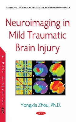
Mild traumatic brain injury (MTBI) is a significant public health problem representing 75% of traumatic brain injury (TBI) cases. There are several advanced techniques available for the investigation of changes in thalamic functions and structures, including a functional MRI using a blood oxygen level-dependent (BOLD) technique as well as diffusion tensor imaging (DTI), including higher-order diffusion kurtosis imaging (DKI) to characterize non-Gaussian water molecular movement with better sensitivity in grey matter, morphological shape, thickness and volumetry as well as a micro-structural level magnetic field correlation (MFC) technique to quantify iron deposition in the brain. Each of these techniques provides important insights into the pathophysiology due to head injury that may not be available in a conventional MRI. For instance, we had used a resting-state (RS) functional connectivity MRI (fcMRI) to investigate the integrity of a default mode network (DMN) with several novel methods, including independent component analysis (ICA) methods in MTBI patients and correlated DMN connectivity changes with neurocognitive tests and clinical symptoms. The authors had further examined thalamic and cortical injuries using a fractional amplitude of low-frequency fluctuations (fALFF) and (fcMRI) based on the resting state (RS) and task-related fMRI in patients. There is an increasing body of literature which suggests that damage to the hypothalamic-hypophysical axis has been under-diagnosed and may account for some of the sequelae following MTBI, but with limited supported imaging finding. The authors had then identified both thalamic and hypothalamic morphology, structure, function and pathway injury using advanced quantitative MRI techniques, including DKI and RS-fMRI to reflect both microstructural and functional connectivity and activity abnormalities in patients. Recent work has also demonstrated significant functional small-world property changes in patients, and suggested decreased global efficiency, possibly due to diffused axonal injury and local network upregulation, including increased thalamo-cortical connectivity. About 20% of patients have persistent post-concussive syndrome (PCS) within 6 months to 1 year after MTBI. The authors find an association between longitudinally regional volume loss in the anterior and isthmus of the cingulum and precuneal regions as well as neurocognitive performance/clinical symptom scores. The brain iron is a promising early imaging biomarker of disease in MTBI, and an MRI is an ideal technique to identify tissue iron in vivo. The quantitative microscopic and macroscopic MFC results demonstrate highly significant differences between patients and controls in both the total and microscopic MFC in the thalamus and significant correlations with an R2* constant, suggesting that the component analysis of MFC is a useful method to study in vivo brain iron after MTBI. And finally, the authors had developed and cross-validated the accuracy of multi-feature algorithms incorporating clinical and imaging metrics to objectively classify patients with MTBI compared to controls. This book will introduce the readers with some perspectives using multi-modality imaging for MTBI disease detection and diagnosis, including several imaging hallmarks: functional, morphological, and microstructural metrics. Various advanced technical developments and the longitudinal monitoring of multi-parametric MRI data will be presented together with some forefront, up-to-date and interesting examples in the MTBI research field. This book will hopefully capture the interests of colleagues in this challenging field and could help convey the technical and developmental information of neuroimaging applications in MTBI.
Yongxia Zhou, PhD Imaging Scientist, University of Southern California and Columbia University, USA
Preface; Characterization of Thalamocortical Association Using the Amplitude & Connectivity of an FMRI in Mild Traumatic Brain Injury; Abnormal Structural & Functional Hypothalamic Connectivity in Mild Traumatic Brain Injury; Default Mode Network Disruption in Mild Traumatic Brain Injury; Small World Property Changes in Mild Traumatic Brain Injury; Assessment of Thalamic Injury Using Structural, Morphological & Functional MRI in Mild Traumatic Brain Injury; Longitudinal Brain Volume Changes in Mild Traumatic Brain Injury Patients; Regional Brain Iron in Mild Traumatic Brain Injury Using a Component Analysis of Magnetic Field Correlation Imaging; Pattern Classification Algorithms Using Multiple MR Imaging Features: An Aid to Identifying Patients with Mild Traumatic Brain Injury; Index.


