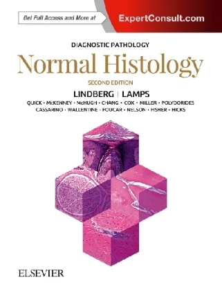
Diagnostic Pathology: Normal Histology
Elsevier - Health Sciences Division (Verlag)
978-0-323-54803-8 (ISBN)
- Titel erscheint in neuer Auflage
- Artikel merken
Zu diesem Buch erhalten Sie kostenlos ein eBook dazu.
This edition incorporates the most recent scientific and technological knowledge in the field to provide a comprehensive overview of all areas of normal histology, including introductory chapters on electron microscopy, immunofluorescence, immunohistochemistry and histochemistry, the cell, and the basic organization of tissues.
With nearly 1,800 outstanding images, this reference is an invaluable diagnostic aid for every practicing pathologist, resident, or fellow.
New to this Edition:
- Thoroughly updated content throughout, with all-new chapters on synovium and histologic artifacts, a thoroughly revised skeletal muscle chapter that now addresses normal histology in the setting of neuromuscular biopsy, and coverage of additional histologic variations that cause diagnostic confusion
- New content on immunohistochemistry; more image examples of newly recognized normal variations, mimics, and pitfalls; and expanded text in many sections for greater clarity and ease of reference
- Expert Consult™ eBook version included with purchase. This enhanced eBook experience allows you to search all of the text, figures, and references from the book on a variety of devices.
Key Features:
- Unparalleled visual coverage with carefully annotated photomicrographs, spectacular gross images, electron micrographs, and medical illustrations
- Time-saving reference features include bulleted text, a variety of test data tables, key facts in each chapter, annotated images, and an extensive index
Matthew R. Lindberg, MD, Assistant Professor of Pathology, Department of Pathology, University of Arkansas for Medical Sciences, Little Rock, Arkansas
INTRODUCTIONTO NORMAL HISTOLOGY AND BASIC HISTOLOGICAL TECHNIQUES
Introduction to the Cell
Introduction to Histology
General Histologic Artifacts
Histochemistry (Special Stains)
Immunohistochemistry of Normal Tissues
Immunofluorescence
Electron Microscopy
INTUGEMENT
Epidermis (Including Keratinocytes and Melanocytes)
Dermis
Adnexal Structures
Nail
MUSCULOSKELETAL SYSTEM
Bone and Cartilage
Synovial Membrane
Connective Tissue
Adipose Tissue
Skeletal Muscle
CIRCULATORY SYSTEM
Heart
Cardiac Valves
Cardiac Conduction System
Arteries
Capillaries, Veins, and Lymphatics
NERVOUSE SYSTEM
Peripheral Nervous System
Central Nervous System
Meninges
Choroid Plexus
HEMATOPOIETIC AND IMMUNE SYSTEMS
Overview of Immune System
Lymph Nodes
Spleen
Bone Marrow
Peripheral Blood
Thymus
HEAD AND NECK
Eye and Ocular Adnexa
Oral Mucosae
Gingivae
Minor Salivary Glands
Major Salivary Glands
Teeth
Tongue
Tonsils/Adenoids
Ear
Nose and Paranasal Sinuses
Pharynx
Larynx
RESPIRATORY SYSTEM
Trachea
Lung
Pleura and Pericardium
BREAST
Breast
TUBULAR GUT AND PERITONEUM
Esophagus
Stomach
Small Intestine
Large Intestine
Appendix
Anus and Anal Canal
Peritoneum
HEPATOBILIARY TRACT AND PANCREAS
Liver
Gallbladder
Extrahepatic Biliary Tract
Vaterian System
Pancreas
BENITOURINARY AND MALE GENITAL TRACT
Kidney
Ureter and Renal Pelvis
Bladder
Urethra
Prostate: Regional Anatomy With Histologic Correlates
Prostate: Benign Glandular and Stromal Histology
Penis
Testis and Associated Excretory Ducts
FEMALE GENITAL TRACT
Vulva
Vagina
Uterus
Fallopian Tube
Ovary
Placenta
ENDOCRINE
Adrenal Gland
Paraganglia
Thyroid
Parathyroid
Pineal Gland
Pituitary
| Erscheinungsdatum | 22.12.2017 |
|---|---|
| Reihe/Serie | Diagnostic Pathology |
| Zusatzinfo | Approx. 1800 illustrations (1800 in full color) |
| Verlagsort | Philadelphia |
| Sprache | englisch |
| Maße | 222 x 281 mm |
| Gewicht | 1540 g |
| Einbandart | gebunden |
| Themenwelt | Studium ► 1. Studienabschnitt (Vorklinik) ► Histologie / Embryologie |
| Studium ► 2. Studienabschnitt (Klinik) ► Pathologie | |
| Schlagworte | diagnostische Pathologie • Histologie • Histologie; Handbuch/Lehrbuch |
| ISBN-10 | 0-323-54803-2 / 0323548032 |
| ISBN-13 | 978-0-323-54803-8 / 9780323548038 |
| Zustand | Neuware |
| Informationen gemäß Produktsicherheitsverordnung (GPSR) | |
| Haben Sie eine Frage zum Produkt? |
aus dem Bereich
