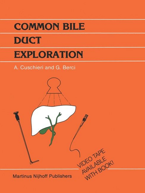
Common Bile Duct Exploration
Kluwer Academic Publishers (Verlag)
978-0-89838-639-4 (ISBN)
- Titel ist leider vergriffen;
keine Neuauflage - Artikel merken
1. Introduction.- 2. Review of existing problems in biliary tract surgery.- 3. Surgical approach and principles.- 1. Introduction.- 2. Prophylactic measures.- 2.1. Infectious complications.- 2.2. Haemorrhagic complications.- 2.3. Renal failure.- 3. Pre-operative biliary decompression in the jaundiced patient.- 4. Operative principles.- 4.1. Surgical access.- 4.2. Patient positioning.- 4.3. Appropriate incision.- 4.4. Illumination of the operating field.- 4.5. Packing.- 4.6. Exposure of relevant anatomy.- 5. Drainage of the supracolic compartment after biliary operations.- 4. Operative cholangiography (in cooperation with J.A. Hamlin and M. Paz-Partlow).- 1. Introduction.- 2. Common bile duct explorations.- 3. Unsuspected stones.- 4. Cannulation techniques.- 5. Initial and/or completion cholangiograms.- 6. Standard technique.- 6.1. Technique and equipment.- 6.2. Patient’s positioning.- 6.3. Scout film.- 6.4. Injected volume.- 6.5. Contrast material.- 6.6. Coordination of exposure.- 6.7. Mobile C-arm fluoroscope.- 7. Fluoro-cholangiography.- 7.1. Easy positioning of the patient.- 7.2. Optimal beam collimation.- 7.3. Shorter exposure time.- 7.4. Automatic exposure control.- 7.5. Minimal technician activity.- 7.6. Control of the exposure sequence.- 7.7. Serial films.- 7.8. Decreased examination time.- 7.9. Indirect radiography.- 8. Anomalies of surgical importance.- 8.1. Short cystic duct.- 8.2. Drainage of cystic duct in the right hepatic duct.- 8.3. Aberrant ducts.- 8.4. Ductal diverticula and choledochocele.- 8.5. The acute or emergency case.- 9. General aspects.- 10. Radiation protection.- 11. The cystic duct.- 12. Cholecysto-cholangiogram.- 13. The choledocho-cholangiogram.- 13.1. Direct needle puncture.- 13.2. Butterfly needle puncture.- 13.3. Special needle clamp.- 13.4. T-tube insertion.- 14. Contact selective cholangiography.- 15. Reason for failure for operative cholangiography.- 15.1. Overfilled ducts.- 15.2. Underfilled ducts.- 15.3. Poor quality films.- 15.4. Improper positioning.- 15.5. Obscured field.- 16. Artifacts.- 17. Complications of operative cholangiography.- 18. Reformed calculi.- 19. Complications of T-tube removal in the post-operative period.- 20. Results of operative cholangiography.- 20.1. Advantages.- 20.2. Disadvantages.- 5. Operative biliary endoscopy (cholangioscopy) (in cooperation with M. Paz-Partlow).- 1. Introduction.- 2. Instrumentation.- 2.1. Accessories.- 3. Technique.- 3.1. Mobilization of the duodenum.- 3.2. Endoscopic appearance.- 3.3. The cystic stump remnant.- 4. Endoscopic anatomy and pathology.- 4.1. Normal findings.- 4.2. Cholangitis.- 4.3. Calculi.- 4.4. Ampullary stenosis.- 4.5. Neoplasms.- 4.6. Miscellaneous.- 5. Repeated cholangioscopy.- 6. Complications.- 7. General aspects.- 7.1. Sterilization.- 7.2. Maintenance.- 8. Evaluation of results.- 9. Conclusions.- 6. Biliary manometry and debimetry.- 1. Introduction.- 2. Usage.- 3. Pharmacolgy of the sphincter of Oddi (SO).- 3.1. Effect of hormones and peptides.- 3.2. Effect of pharmacological agents.- 4. Biliary pressure indices.- 4.1. Resting (initial, interdigestive) pressure.- 4.2. Passage (yield, opening) pressure.- 4.3. Filling pressure curves.- 4.4. Residual pressure.- 4.5. Flow rate (debimetry).- 4.6. Incremental pressure and recovery time.- 5. Dynamic (transducer) manometry.- 5.1. Endoscopic sphincter zone activity.- 5.2. Technique of operative biliary manometry.- 6. Disorders of the sphincter of Oddi.- 6.1. Iatrogenic stricture.- 6.2. Papillitis/Oedema.- 6.3. Papillary stenosis (choledocho-duodenal junctional stenosis).- 6.4. Functional disorders.- 7. Exploration of the common bile duct.- 1. Introduction.- 2. Technique of CBD exploration.- 2.1. Mobilization of duodenum and head of pancreas.- 2.2. Exposure of the CBD.- 2.3. Choledochotomy.- 2.4. Cholangioscopy.- 2.5. Additional procedures.- 2.6. Insertion of T-tube.- 2.7. Closure of choledochotomy wound.- 3. Trans-duodenal exploration od CBD.- 4. Intra-hepatic calculi.- 5. Assessment of terminal end of the CBD and sphincteric region.- 6. Post-operative removal of T-tube.- 7. Conclusion.- 8. Postoperative removal of retained stones through the T-Tube tract (in cooperation with J.A. Hamlin).- 1. Introduction.- 2. Stone extraction via the T-tube.- 3. Endoscopic method.- 4. Preparation for stone extraction.- 5. Technique.- 6. Results.- 7. Complications.- 8. Discussion.- Index of Subjects.
| Reihe/Serie | Developments in Surgery ; 6 |
|---|---|
| Zusatzinfo | VIII, 100 p. |
| Verlagsort | Dordrecht |
| Sprache | englisch |
| Gewicht | 600 g |
| Themenwelt | Medizin / Pharmazie ► Medizinische Fachgebiete ► Chirurgie |
| Medizinische Fachgebiete ► Innere Medizin ► Gastroenterologie | |
| Medizinische Fachgebiete ► Innere Medizin ► Hepatologie | |
| ISBN-10 | 0-89838-639-X / 089838639X |
| ISBN-13 | 978-0-89838-639-4 / 9780898386394 |
| Zustand | Neuware |
| Haben Sie eine Frage zum Produkt? |
aus dem Bereich


