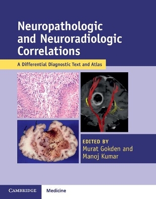
Neuropathologic and Neuroradiologic Correlations
Cambridge University Press
978-1-107-56725-2 (ISBN)
Neuroscience is an evolving, complex, and multidisciplinary field of medicine. Neuroradiologists and neuropathologists are an important part of the multidisciplinary team, in addition to neurosurgeons, neurologists, and other disciplines. Featuring over nine hundred images, this practical textbook and atlas combines the specialties of neuroradiology and neuropathology, providing an extensive understanding of the disease process. It offers a comprehensive review of the nervous system diseases, including eye, skeletal muscle, and bone and soft tissue diseases. Topics are covered in chapters arranged by region, allowing for quick reference of conditions such as brain tumors, spinal cord diseases, or congenital malformations. Introductory chapters on pathologic and radiologic techniques are also featured, enabling specialists of both areas to familiarize themselves with the other's subject. Packaged with a password to give the user online access to all the text and images, this is a must-have resource for comprehensive and accurate diagnosis.
Murat Gokden is Professor of Pathology and Neuropathology in the Department of Pathology, University of Arkansas for Medical Sciences and Arkansas Children's Hospital, Little Rock. Manoj Kumar is Associate Professor of Neuroradiology in the Department of Radiology, University of Arkansas for Medical Sciences, Little Rock.
List of contributors; Preface; 1. Radiologic techniques Manoj Kumar; 2. Pathologic techniques Theodore Friedman and Mahtab Tehrani; 3. Meningeal mass lesions Mahlon D. Johnson and Ali Hussain; 4. Diffuse leptomeningeal and dural lesions Ali Hussain and Mahlon D. Johnson; 5. Sellar and suprasellar region Beatriz Lopes and Prashant Raghavan; 6. Pineal region Melissa Gener, Stephen Kralik and Eyas Hattab; 7. Mass effect and edema Bret Evers, Travis Danielsen and Manoj Kumar; 8. Cerebral mass lesions Douglas C. Miller and Girish M. Fatterpekar; 9. Cerebral atrophy Robert E. Mrak and Edgardo J. C. Angtuaco; 10. Ventricular system Bruce C. Gilbert, Suash Sharma, Ramon Figueroa and Amyn M Rojiani; 11. White matter Suash Sharma, Brandi Villarreal, John Edry, Reed Murtagh and Amyn M. Rojiani; 12. Cerebellum and brainstem mass lesions Raghu H. Ramakrishnaiah, Veena Rajaram and Charles M. Glasier; 13. Malformations Veena Rajaram and Korgun Koral; 14. Cerebellopontine angle Rohan S. Samant and Murat Gokden; 15. Spinal cord Sumit Singh and S. Humayun Gultekin; 16. Bone and soft tissues Roopa Ram, Kedar Jambhekar and Robin Elliott; 17. Peripheral nervous system Shivani Ahlawat, Laura M. Fayad and Fausto Rodriguez; 18. Skeletal muscle S. Humayun Gultekin and Brooke Beckett; 19. Ophthalmic diseases Anat O. Stemmer-Rachamimov, Nora Laver, Declan McGuone, Baiju Shah, Gene M. Weinstein and Harprit Singh Bedi; Index.
| Erscheint lt. Verlag | 29.6.2017 |
|---|---|
| Zusatzinfo | 49 Tables, black and white; 373 Halftones, color; 581 Halftones, black and white |
| Verlagsort | Cambridge |
| Sprache | englisch |
| Maße | 225 x 283 mm |
| Gewicht | 1990 g |
| Themenwelt | Medizin / Pharmazie ► Medizinische Fachgebiete ► Neurologie |
| Medizin / Pharmazie ► Medizinische Fachgebiete ► Radiologie / Bildgebende Verfahren | |
| Studium ► 2. Studienabschnitt (Klinik) ► Pathologie | |
| ISBN-10 | 1-107-56725-4 / 1107567254 |
| ISBN-13 | 978-1-107-56725-2 / 9781107567252 |
| Zustand | Neuware |
| Informationen gemäß Produktsicherheitsverordnung (GPSR) | |
| Haben Sie eine Frage zum Produkt? |
aus dem Bereich

