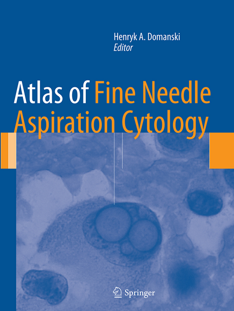
Atlas of Fine Needle Aspiration Cytology
Seiten
2016
|
Softcover reprint of the original 1st ed. 2014
Springer London Ltd (Verlag)
978-1-4471-6954-3 (ISBN)
Springer London Ltd (Verlag)
978-1-4471-6954-3 (ISBN)
The Atlas covers all diagnostic areas where fine needle aspiration cytology is used, including palpable lesions and lesions sampled using radiological methods, and correlations with ancillary examinations. Thouroughly illustrated, with abundant color images.
This book covers all of the diagnostic areas where FNAC is used today. This includes palpable lesions and lesions sampled using various radiological methods, and correlations with ancillary examinations detailed on an entity-by-entity basis. As well as being a complete atlas of the facts and findings important to FNAC, this atlas is a guide to diagnostic methods that optimize health care. The interaction of the cytologist or cytopathologist with other specialists (radiologists, oncologists and surgeons) involved in the diagnosis and treatment of patients with suspicious mass lesions is emphasized and illustrated throughout.
With contributions from experts in the field internationally and abundant colour images Atlas of Fine Needle Aspiration Cytology provides a comprehensive and up-to-date guide to FNAC for pathologists, cytopathologists, radiologists, oncologists, surgeons and others involved in the diagnosis and treatment of patients with suspicious mass lesions.
Editor Henryk A. Domanski Department of Pathology and Cytology, Lund University Hospital, Sweden
Introduction.- Image-Guided Fine Needle Aspiration Cytology.- Breast.- Head and Neck: Salivary Glands.- Thyroid.- Lung.- Mediastinum and Endobronchial Ultrasound-Guided Transbronchial Needle Aspiration.- Lymph Nodes.- Spleen.- Liver.- Pancreas.- Kidney and Adrenal Gland.- Soft Tissue.- Skin and Subcutis.- Bone.- Pediatric Tumours.- Orbit and Ocular Adnexa.
| Erscheinungsdatum | 28.02.2017 |
|---|---|
| Zusatzinfo | 600 Illustrations, color; 11 Illustrations, black and white; XII, 572 p. 611 illus., 600 illus. in color. |
| Verlagsort | England |
| Sprache | englisch |
| Maße | 210 x 279 mm |
| Themenwelt | Medizin / Pharmazie ► Medizinische Fachgebiete |
| Studium ► 1. Studienabschnitt (Vorklinik) ► Histologie / Embryologie | |
| Studium ► 2. Studienabschnitt (Klinik) ► Anamnese / Körperliche Untersuchung | |
| Studium ► 2. Studienabschnitt (Klinik) ► Pathologie | |
| ISBN-10 | 1-4471-6954-9 / 1447169549 |
| ISBN-13 | 978-1-4471-6954-3 / 9781447169543 |
| Zustand | Neuware |
| Informationen gemäß Produktsicherheitsverordnung (GPSR) | |
| Haben Sie eine Frage zum Produkt? |
Mehr entdecken
aus dem Bereich
aus dem Bereich
Zytologie, Histologie und mikroskopische Anatomie
Buch | Hardcover (2022)
Urban & Fischer in Elsevier (Verlag)
54,00 €


