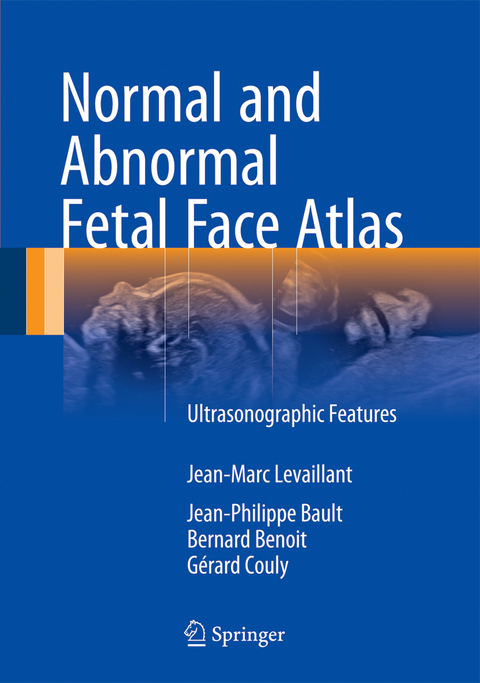
Normal and Abnormal Fetal Face Atlas
Springer International Publishing (Verlag)
978-3-319-43768-2 (ISBN)
The overall examination of a newborn's face offers a rich source of information and can guide the general examination. The same applies in the context of fetal ultrasound examination. The analytical study of the fetal face not only makes it possible to screen for anomalies related to the face itself, but also yields valuable insights into the brain, the limbs, and the heart. In addition, it allows ultra-sonographers to unravel the puzzle of fetal dimorphism.
Written in a pedagogical style, the book guides walks the reader through the diagnostic reasoning process step by step.
The authors are pioneers in this field and teach in various university and master's degree ultrasound programs. Their aim is to share with readers their diagnostic approaches and their knowledge and passion for 2D and 3D ultrasound techniques. Each chapter includes algorithms, biometry curves, and simple guidelines that allow users to go "from sign to syndrome". The first chapter, which focuses on innovative embryology adapted to the needs of ultra-sonographers, was written by Gérard Couly, a maxilla-facial surgeon and the founding father of the specialty>
Jean-Marc Levaillant is a gynecologist-obstetrician specialized in fetal medicine at the Bicetre Prenatal Diagnosis Center and the Créteil Center for Women and Fetal Imaging, in France. Jean-Philippe Bault is a gynecologist-obstetrician specialized in fetal medicine at the Poissy Prenatal Diagnosis Center, France. Bernard Benoit is a gynecologist-obstetrician specialized in fetal medicine at the Princess GRACE Hospital, Monaco. Jean-Marc Levaillant, Bernard Benoit, and Jean-Philippe Bault are all three pioneers in the field and teach various university and master's degree ultrasound programs, and run the Kremlin-Bicêtre 3D Ultrasound School. They share with readers their diagnostic approach and their passion for 2D and 3D ultrasound. Gérard Couly, a maxilla-facial surgeon, is the founding father of this specialty and kindly wrote the first chapter on innovative embryology adapted to ultrasonographers. It is of rare intelligence and will remain a reference for understanding ultrasonography of the face.
Foreword.- Preface.- Introduction.- Key phases in cranial-facial development.- Cellular phenotypes created by the neural crest.- Summary.- Chapter 1. Normal Face<.- Chapter 2. Clefts and Pierre-Robin Syndrome.- Chapter 3. Dysmorphism.- Chapter 4. Upper level pathologies.- Chapter 5. Mid-level pathologies.- Chapter 6. Lower level pathologies.- Chapter 7. Multiple-level pathologies.- Chapter 8. Additional syndromes.- Chapter 9. Facial tumors.- Chapter 10. The eye.- Chapter 11. Key points on eye pathologies.- Chapter 12. Curves and biometrics
| Erscheinungsdatum | 29.03.2017 |
|---|---|
| Zusatzinfo | XI, 223 p. 280 illus., 239 illus. in color. |
| Verlagsort | Cham |
| Sprache | englisch |
| Maße | 178 x 254 mm |
| Themenwelt | Medizinische Fachgebiete ► Radiologie / Bildgebende Verfahren ► Radiologie |
| Schlagworte | 2D and 3D ultrasound • anatomy • diagnostic radiology • Facial clefts • Facial dysmorphism • Facial embryology • Fetal Medicine • Medicine • obstetrics/perinatology • ultrasonography • Ultrasound |
| ISBN-10 | 3-319-43768-2 / 3319437682 |
| ISBN-13 | 978-3-319-43768-2 / 9783319437682 |
| Zustand | Neuware |
| Haben Sie eine Frage zum Produkt? |
aus dem Bereich


