Compendium of Histology
Springer International Publishing (Verlag)
978-3-319-41871-1 (ISBN)
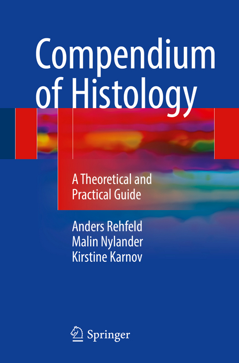
This book has been designed to help medical students succeed with their histology classes, while using less time on studying the curriculum. The book can both be used on its own or as a supplement to the classical full-curriculum textbooks normally used by the students for their histology classes. Covering the same curriculum as the classical textbooks, from basic tissue histology to the histology of specific organs, this book is formatted and organized in a much simpler and intuitive way. Almost all text is formatted in bullets or put into structured tables. This makes it quick and easy to digest, helping the student get a good overview of the curriculum. It is easy to locate specific information in the text, such as the size of cellular structures etc. Additionally, each chapter includes simplified illustrations of various histological features. The aim of the book is to be used to quickly brush up on the curriculum, e.g. before a class or an exam. Additionally, the book includes guides to distinguish between the different histological tissues and organs that can be presented to students microscopically, e.g. during a histology spot test. This guide lists the specific characteristics of the different histological specimens and also describes how to distinguish a specimen from other similar specimens. For each histological specimen, a simplified drawing and a photomicrograph of the specimen, is presented to help the student recognize the important characteristics in the microscope. Lastly, the book contains multiple "memo boxes" in which parts of the curriculum are presented as easy-to-remember mnemonics.
Anders Rehfeld Anders Rehfeld finished his MD from the University of Copenhagen in 2014 and earned his PhD in male reproductive biology from the University of Copenhagen in 2017. He began teaching histology during the 3rd year of his medical studies and has since taught 19 classes of histology of tissues and basic cell biology at the University of Copenhagen. From the beginning of his teaching period, he has been eager to help his students comprehend the large curriculum in a smarter and faster way, and in 2012 his efforts culminated with the publishing of the first compendium on histology Histologi kompendium (in Danish) by theauthors. Malin NylanderMalin Nylander graduated as a MD from the University of Copenhagen in 2012 and earned her PhD in gynecological endocrinology from the University of Copenhagen in 2017. With a great interest in human biology, she started teaching anatomy and histology halfway through medical school and has taught several classes of anatomy, histology, and human biology at the University of Copenhagen. Using a systematic approach combined with simple, schematic drawings on the black board, she tried to make the, at times, complicated curriculum comprehensible to all—something she implemented in the first compendium on histology Histologi kompendium (in Danish) published by the authors in 2012. Kirstine Karnov In 2013 Kirstine Karnov obtained her MD from the University of Copenhagen. She is in residency to become an otorhinolaryngologist and is currently doing a PhD on oral cancer. She began teaching histology during the 3rd year of her medical studies and has taught multiple classes of anatomy, dissection, and histology at the University of Copenhagen during a period of 5 years. Teaching has been one of her most enjoyable and learning experiences in her professional career, and the fun in teaching culminated in 2012 when the first compendium on histology Histologi kompendium (in Danish) was published by the authors.
From cells to tissues.- Histological methods.- The cytoplasm.- The nucleus.- Epithelial tissue.- Glandular epithelium and glands.- Connective tissue.- Cartilage.- Bone tissue.- Bone marrow.- Adipose tissue.- Blood.- Muscle tissue.- Nerve tissue.- The Musculoskeletal system.- The Nervous system.- The Cardiovascular system.- The Respiratory system.- The Immune system & the lymphatic organs.- The integumentary system.- The digestive system I: The alimentary canal.- The digestive system II: The associated organs.- The urinary system.- The Endocrine system.- The female reproductive system.- The male reproductive system.- The breast (mamma).- Eye.- Ear.
"This book is an ideal companion for medical students learning histology and for revision prior to examinations. With an excellent and concise overview of the main organ systems and with no extraneous material, the book perfectly achieves its set purpose. ... this book would be an expense-free venture for students. It would be invaluable to those wishing to establish a foundation in histology and those individuals who have a special interest in pathology." (Sue Sritharan, AMSJ Australian Medical Student Journal, Vol. 10 (1), 2020)
| Erscheinungsdatum | 06.10.2017 |
|---|---|
| Zusatzinfo | XIX, 675 p. 137 illus. in color. |
| Verlagsort | Cham |
| Sprache | englisch |
| Maße | 155 x 235 mm |
| Themenwelt | Medizin / Pharmazie ► Medizinische Fachgebiete |
| Studium ► 2. Studienabschnitt (Klinik) ► Pathologie | |
| Schlagworte | Connective Tissue • Epithelial tissue • Histological methods • Histology • Medicine • Microscopic Anatomy • Pathology • Specialized histology |
| ISBN-10 | 3-319-41871-8 / 3319418718 |
| ISBN-13 | 978-3-319-41871-1 / 9783319418711 |
| Zustand | Neuware |
| Haben Sie eine Frage zum Produkt? |
aus dem Bereich


