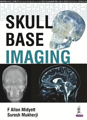
Skull Base Imaging
Jaypee Brothers Medical Publishers (Verlag)
978-93-85999-31-4 (ISBN)
- Titel wird leider nicht erscheinen
- Artikel merken
This book is a complete guide to skull base imaging covering all current techniques including CT, MRI, Ultrasound, Angiography, CT Cisternography and Plain Film Radiography.
Divided into eight sections, each part covers imaging techniques for a different region of the skull base, including cerebellopontine, anterior, posterior and middle cranial fossa, the pituitary region, craniovertebral junction and more. The final chapters discuss inflammatory disorders and sarcomas.
The book includes the very latest techniques including diffusion weighted imaging (DWI) and FLAIR. Each chapter features key points, clues, incidence and location, as well as detail on presentation, epidemiology, pathology, staging, differential diagnosis, treatment and prognosis.
Authored by recognised experts from Washington and Michigan, this comprehensive guide is enhanced by radiological images and illustrations.
Key Points
Complete guide to skull base imaging
Covers all regions of the skull base
Includes latest techniques including DWI and FLAIR
US-based, expert author team
F Allan Midyett MD Neuroradiologist, Georgia, Washington, USA Suresh Mukherji MD MBA FACR Professor & Chair, Department of Radiology, Michigan State University, USA
Section I: Pituitary Region
RCC, Rathke's Cleft Cyst
Pituitary Adenoma
Craniopharyngioma
EN, Ectopic Neurohypohysis
HH,Hypothalamic Hamartoma
Neurohypophyseal Sarcoidosis
Arachnoid Cyst
Langerhans Cell Histiocytosis
Section II: Cerebellopontine Angle
Epidermoid
CPA Meningioma
Vestibular Schwannomas
Arachnoid Cyst
Section III: Anterior Cranial Fossa
Anterior Fossa Meningioma
IP, Inverted Papilloma
JNA, Juvenile Nasal Angiofibroma
NEC, Neuroendocrine Carcinoma
NFE, Nasofrontal Encephalocele
SNM, Sinonasal Melanoma
Nasal Dermoid
Nasal Glioma
Paranasal Sinus Mucocele
Osteoma
SNUC
Skull Base Fx/CSF Leaks
Adenoid Cystic Carcinoma
Paranasal Sinus Cancer
Section IV: Middle Cranial Fossa
Jugular Foramen Paraganglioma
Clivus Chordoma
LM, Lymphatic Malformations
AFS, Allergic Fungal Sinusitis
MCF 5, Basal Encephalocele
Cavernous Sinus Aneurysm
Gradenigo's Syndrome
Cavernous Sinus Thrombosis
Cavernous Sinus Lymphoma
Aneurysmal Bone Cyst
Cholesteatoma
Giant Aneurysm
Giant Cell Tumor
Giant Osteoma
Plasmacytoma/ Myeloma
Metastatic Prostate
Choclear Implants
Petrous Apex Granuloma
Gradenigo's Syndrome
Section V: Craniovertebral Junction
AOJ Dislocation
Chiari Malformation
CVJ Lymphoma
Congenital Abnormalities (other)
Paget Disease
Section VI: Posterior Cranial Fossa
PFAC Posterior Fossa Arachnoid Cyst
Meningioma
Hemangioblastoma/Hemangiopericytoma
Metastasis
Plasmacytoma medulloblastoma
Section VII: Inflammatory
Skull Base Osteomyelitis
Neurosyphilis
Sarcoidosis
Danger Space Abscess
Wegeners
Cistercercosis
Section VIII: Sarcomas
Chondrosarcoma
Ewings Sarcoma
Osteosarcoma
Fibrosarcoma
Undifferentiated Sarcoma
Rhabdomyosarcoma
Synovial Sarcoma
Angiosarcoma
| Erscheinungsdatum | 01.02.2017 |
|---|---|
| Verlagsort | New Delhi |
| Sprache | englisch |
| Maße | 216 x 279 mm |
| Gewicht | 1300 g |
| Themenwelt | Medizinische Fachgebiete ► Chirurgie ► Neurochirurgie |
| Medizin / Pharmazie ► Medizinische Fachgebiete ► HNO-Heilkunde | |
| Medizin / Pharmazie ► Medizinische Fachgebiete ► Orthopädie | |
| Medizin / Pharmazie ► Medizinische Fachgebiete ► Radiologie / Bildgebende Verfahren | |
| ISBN-10 | 93-85999-31-1 / 9385999311 |
| ISBN-13 | 978-93-85999-31-4 / 9789385999314 |
| Zustand | Neuware |
| Haben Sie eine Frage zum Produkt? |
aus dem Bereich


