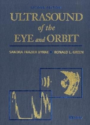
Ultrasound of the Eye and Orbit
Mosby (Verlag)
978-0-323-01207-2 (ISBN)
- Titel ist leider vergriffen;
keine Neuauflage - Artikel merken
Completely revised and updated, the new 2nd Edition of the classic text in the field encompasses all of the latest information on ophthalmic ultrasound. Readers will find in-depth coverage of new technology, new detailed descriptions of examination techniques, and the latest uses of ultrasound in intraocular and orbital lesions. All chapters have been expanded to include new disease entities, and new illustrations have been added for greater clarity.
1.Physics and Instrumentation History Physics Instrumentation The Globe Introduction Indications 2.Examination Techniques for the Eye Positioning the Patient B-scan Examination Techniques Basic Screening Examination Special Examination Techniques Anterior Segment Evaluation: Immersion Technique Evaluation of the Lens Evaluation of the Pupil Pediatric Examination Documentation of Findings 3.Vitreoretinal Disease Vitreous Body Retina Retinal Pigment Epithelium Macula Choroid Ciliary Body Sclera 4.Trauma and Postsurgical Findings Blunt Trauma Penetrating Trauma Surgical Trauma (Complications) Postsurgical Findings 5.Intraocular Tumors Detection of Lesions Ocular Melanoma Other Tumors of the Uvea, Retina, and RPE Other Lesions Simulating Melanoma Anterior Segment Tumors Retinoblastoma Other Lesions Associated with Leukokoria 6.Inflammatory Diseases of the Eye Endophthalmitis Noninfectious Uveitis/Vitritis Scleritis Inflammatory Conditions of the Choroid Miscellaneous Conditions Associated with Choroidal Inflammation of Choroid, Retina, and RPE 7.Glaucoma Optic Disc Congenital Glaucoma Angle Closure Glaucoma Secondary Glaucoma Normal-Tension Glaucoma Complications of Glaucoma Surgery Glaucoma Filtering Implant Devices 8.Ultrasound Biomicroscopy of the Eye Theoretical Consideration and Development of the Ultrasound Biomicroscope Clinical Use of Ultrasound Biomicroscopy Ultrasound Biomicroscopy in Ocular Disease Summary and Future Directions 9.Three-Dimensional Ultrasound of the Eye Introduction Instrumentation Intraocular Tumors Vitreoretinal Disease Advantages and Limitations of Ophthalmic 3D Ultrasound Future Directions and the Internet 10.Axial Eye Length Measurements (A-Scan Biometry) Standard Axial Eye Length Dimensions Instrumentation Instrument Settings Examination Procedures for A-Scan Biometry Troubleshooting Minimizing Errors IOL Calculations Unanticipated Postoperative Refractive Errors Diagnostic Uses of Axial Eye Length Measurements Cleaning and Calibration of Biometry Instruments The Orbit Introduction Indications 11.Examination Techniques for the Orbit Positioning the Patient B-scan Techniques Basic Examination for Lesion Detection Special Examination Techniques for Lesion Differentiation Quantitative Echography Kinetic Echography 12.Orbital Tumors Pseudotumor and Lymphoma (Lymphoproliferative Diseases) Primary Orbital Tumors Metastatic and Secondary Tumors Lacrimal System Disorders Cystic Lesions 13.Vascular Lesions Vascular Neoplasms Vascular Malformations 14.Color Doppler Imaging of the Eye and Orbit Introduction Color-Duplex Scanning Physical Background and Imaging Devices Clinical Application of CDI Ophthalmic Examination of the Eye and Orbit Retinal, Retinal Vascular, and Other Vascular Diseases of the Eye Intraocular Tumors Orbital Disorders Safety Considerations 15.Extraocular Muscles Examination Techniques for Rectus Muscles Evaluation of Individual Muscles Extraocular Muscle Disorders 16.Optic Nerve Retrobulbar Optic Nerve Retrobulbar Optic Nerve Disorders Optic Disc 17.Trauma and Periorbital Disease Trauma Periorbital Disease 18.Artifacts Multiple Signals (Reverberations) Shadowing Enhancement Perpendicular Sound Beam Incidence Baums Bumps Glossary Appendices Index
| Erscheint lt. Verlag | 23.5.2002 |
|---|---|
| Zusatzinfo | 1857 ills |
| Verlagsort | St Louis |
| Sprache | englisch |
| Maße | 216 x 279 mm |
| Gewicht | 1805 g |
| Themenwelt | Medizin / Pharmazie ► Medizinische Fachgebiete ► Augenheilkunde |
| Medizinische Fachgebiete ► Radiologie / Bildgebende Verfahren ► Sonographie / Echokardiographie | |
| ISBN-10 | 0-323-01207-8 / 0323012078 |
| ISBN-13 | 978-0-323-01207-2 / 9780323012072 |
| Zustand | Neuware |
| Haben Sie eine Frage zum Produkt? |
aus dem Bereich


