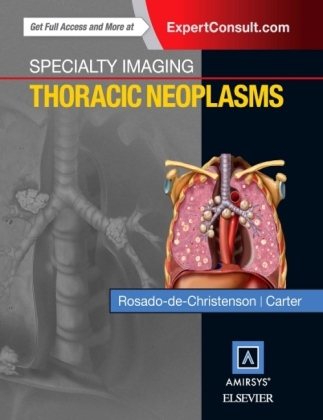
Specialty Imaging: Thoracic Neoplasms
Seiten
2015
Elsevier - Health Sciences Division (Verlag)
978-0-323-37706-5 (ISBN)
Elsevier - Health Sciences Division (Verlag)
978-0-323-37706-5 (ISBN)
- Titel ist leider vergriffen;
keine Neuauflage - Artikel merken
Part of the highly regarded Specialty Imaging series, this unique title by Dr. Melissa L. Rosado-de-Christenson clearly presents the imaging features of all thoracic neoplasms (including those affecting the cardiovascular system) as well as staging of malignancies and patterns of metastatic spread in a single, convenient volume. An easy-to-read bulleted format and state-of-the-art imaging examples guide you step by step through every aspect of the field, including invasive diagnostic and therapeutic procedures. This book is an ideal resource for radiologists, pulmonary medicine physicians, thoracic surgeons, thoracic oncologists, and radiation oncologists - anyone who must distinguish lung cancer and thoracic metastases from less common malignant and benign neoplasms.
Superb illustrations highlight comprehensive coverage of imaging manifestations of all benign and malignant thoracic neoplasms, including lesions in the lung, mediastinum, thymus, esophagus, cardiovascular system, pleura, and chest wall
Introductory chapters discuss the various imaging modalities used in diagnosing and evaluating thoracic neoplasms, up-to-date imaging terminology, and the imaging signs that suggest neoplasia
Thorough coverage of lung cancer offers authoritative guidance on screening, specific manifestations of various cell types, issues of staging, various tissue-sampling methods, missed lung cancer, lung cancer mimics, and imaging follow-up of treated lung cancer
High-quality images and succinct text depict patterns of thoracic metastatic spread of several important malignancies, navigational bronchoscopy and image-guided biopsy, imaging manifestations of treated patients, and other key topics
A time-saving bulleted format distills essential information for fast and easy comprehension
Professor, University of Missouri-Kansas City School of Medicine, Department of Radiology, Kansas city, MO, United States.
| Reihe/Serie | Specialty Imaging |
|---|---|
| Zusatzinfo | Approx. 1500 illustrations (1500 in full color); Illustrations |
| Verlagsort | Philadelphia |
| Sprache | englisch |
| Maße | 222 x 281 mm |
| Gewicht | 2070 g |
| Themenwelt | Medizin / Pharmazie ► Medizinische Fachgebiete ► Onkologie |
| Medizinische Fachgebiete ► Radiologie / Bildgebende Verfahren ► Radiologie | |
| ISBN-10 | 0-323-37706-8 / 0323377068 |
| ISBN-13 | 978-0-323-37706-5 / 9780323377065 |
| Zustand | Neuware |
| Haben Sie eine Frage zum Produkt? |
Mehr entdecken
aus dem Bereich
aus dem Bereich
Buch (2023)
Thieme (Verlag)
190,00 €


