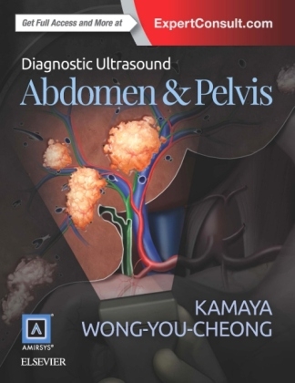
Diagnostic Ultrasound: Abdomen and Pelvis
Elsevier - Health Sciences Division (Verlag)
978-0-323-37643-3 (ISBN)
- Titel erscheint in neuer Auflage
- Artikel merken
Coverage of new topics including liver transplantation, bowel ultrasound, and other various abdominal and pelvic entities
Detailed anatomy section shows transducer placement in association with imaging, with a robust collection of CT/MR correlations
Time-saving reference features include succinct and bulleted text, a variety of test data tables, key facts in each chapter, annotated images, and an extensive index
Expert Consult eBook version included with purchase. This enhanced eBook experience allows you to search all of the text, figures, images, and references from the book on a variety of devices.
Aya Kamaya, MD is an Associate Professor of Radiology at Stanford University. She is the Director of Ultrasound and Body Imaging Fellowship Director. Her clinical focuses are Oncologic Imaging, Hepatobiliary and Pancreatic Imaging, Urogenital Imaging, Gynecological Imaging, Thyroid Ultrasound, Ultrasound, and Diagnostic Radiology. Kamaya is the co-lead author from Diagnostic Ultrasound: Abdomen and Pelvis, 1e. Jade J. Wong-You-Cheong, MD is a Professor of Diagnostic Radiology and Nuclear Medicine at the University of Maryland. She also has a secondary appointment in Surgery and is the Vice Chair of Quality and Patient Safety. Wong is the co-lead author from Diagnostic Ultrasound: Abdomen and Pelvis, 1e.
PART I: ANATOMY
Abdomen
1 Liver
2 Biliary System
3 Spleen
4 Pancreas
5 Kidneys
6 Adrenals
7 Bowel
8 Abdominal Lymph Nodes
9 Peritoneal Spaces and Structures
10 Abdominal Wall
Pelvis
11 Ureters and Bladder
12 Prostate
13 Testes
14 Uterus
15 Cervix
16 Vagina
17 Ovaries
PART II: DIAGNOSES
Liver
Introduction and Overview
18 Approach to Hepatic Sonography
Diffuse Parenchymal Disease
19 Acute Hepatitis
20 Hepatic Cirrhosis
21 Hepatic Steatosis
22 Hepatic Schistosomiasis
23 Venoocclusive Disease
Cyst and Cyst-Like Lesions
24 Hepatic Cyst
25 Biliary Hamartoma
26 Caroli Disease
27 Biloma
28 Biliary Cystadenoma/Carcinoma
29 Pyogenic Hepatic Abscess
30 Amebic Hepatic Abscess
31 Hepatic Echinococcus Cyst
32 Hepatic Diffuse Microabscesses
33 Peribiliary Cyst
34 Hepatic Foregut Cyst
Focal Solid Masses
35 Hepatic Cavernous Hemangioma
36 Focal Nodular Hyperplasia
37 Hepatic Adenoma
38 Hepatocellular Carcinoma
39 Hepatic Metastases
40 Hepatic Lymphoma
Vascular Conditions
41 Portal Hypertension
42 TIPS Shunts
43 Portal Vein Occlusion
44 Budd-Chiari Syndrome
45 Portal Vein Gas
Liver Transplants
46 Hepatic Artery Stenosis/Occlusion
47 Portal Vein Stenosis/Occlusion
48 Venous Stenosis
49 Common Bile Duct Stricture
Biliary System
Introduction and Overview
50 Approach to Biliary Sonography
Gallstones and Mimics
51 Cholelithiasis
52 Echogenic Bile
53 Gallbladder Cholesterol Polyp
Gallbladder Wall Pathology
54 Acute Calculous Cholecystitis
55 Acute Acalculous Cholecystitis
56 Chronic Cholecystitis
57 Xanthogranulomatous Cholecystitis
58 Porcelain Gallbladder
59 Hyperplastic Cholecystosis
60 Gallbladder Carcinoma
Ductal Pathology
61 Biliary Ductal Dilatation
62 Choledochal Cyst
63 Choledocholithiasis
64 Biliary Ductal Gas
65 Cholangiocarcinoma
66 Ascending Cholangitis
67 Recurrent Pyogenic Cholangitis
68 AIDS-Related Cholangiopathy
Pancreas
Introduction and Overview
69 Approach to Pancreatic Sonography
Pancreatitis
70 Pancreatitis, Acute
71 Pancreatic Pseudocyst
72 Pancreatitis, Chronic
Simple Cysts and Cystic Neoplasms
73 Mucinous Cystic Pancreatic Tumor
74 Serous Cystadenoma of Pancreas
75 IPMN
Solid-Appearing Pancreatic Neoplasms
76 Pancreatic Ductal Carcinoma
77 Pancreatic Neuroendocrine Tumors
78 Solid Pseudopapillary Neoplasm
Spleen
Introduction and Overview
79 Approach to Splenic Sonography
Splenic Lesions
80 Splenomegaly
81 Splenic Cyst
82 Splenic Tumors
83 Splenic Infarct
Urinary Tract
Introduction and Overview
84 Approach to Urinary Tract Sonography
Normal Variants and Pseudolesions
85 Column of Bertin, Kidney
86 Renal Junction Line
87 Renal Ectopia
88 Horseshoe Kidney
89 Ureteral Duplication
90 Ureteral Ectopia
91 Ureteropelvic Junction Obstruction
Calculi and Calcinosis
92 Urolithiasis
93 Nephrocalcinosis
94 Hydronephrosis
Cysts and Cystic Disorders
95 Simple Renal Cyst
96 Complex Renal Cyst
97 Cystic Disease of Dialysis
98 Lithium Microcysts
99 Multilocular Cystic Nephroma
Urinary Tract Infection
100 Acute Pyelonephritis
101 Renal Abscess
102 Emphysematous Pyelonephritis
103 Pyonephrosis
104 Xanthogranulomatous Pyelonephritis
105 Tuberculosis, Urinary Tract
Solid Renal Neoplasms
106 Renal Cell Carcinoma
107 Renal Metastases
108 Renal Angiomyolipoma
109 Transitional Cell Carcinoma
110 Renal Lymphoma
Vascular Conditions
111 Renal Artery Stenosis
112 Renal Vein Thrombosis
113 Renal Infarct
114 Perinephric Hematoma
Prostate
115 Prostatic Hypertrophy
116 Prostatic Carcinoma
Bladder
117 Bladder Carcinoma
118 Ureterocele
119 Bladder Diverticulum
120 Bladder Calculi
121 Schistosomiasis, Bladder
Kidney
Introduction and Overview
122 Approach to Sonographic Features of Renal Allografts
Renal Transplant Complications
123 Allograft Hydronephrosis
124 Perigraft Fluid Collections
125 Allograft Rejection
126 Renal Transplant Vascular Disorders
127 AV Fistula/Pseudoaneurysm
Adrenal Gland
127 Adrenal Hemorrhage
128 Myelolipoma
129 Adrenal Adenoma
130 Adrenal Cysts
131 Pheochromocytoma
132 Adrenal Carcinoma
Abdominal Wall/Peritoneal Cavity
133 Abdominal Wall Hernia
134 Groin Hernia
135 Ascites
136 Peritoneal Carcinomatosis
137 Peritoneal Space Abscess
Bowel
138 Appendicitis
139 Appendiceal Mucocele
140 Intussusception
141 Epiploic Appendagitis
142 Diverticulitis
143 Crohn Disease
144 Bowel Malignancy
Scrotum
Introduction and Overview
145 Approach to Scrotal Sonography
Scrotal Lesions
146 Testicular Germ Cell Tumors
147 Gonadal Stromal Tumors, Testis
148 Testicular Lymphoma/Leukemia
149 Epidermoid Cyst
150 Tubular Ectasia of Rete Testes
151 Testicular Microlithiasis
152 Testicular Torsion/Infarction
153 Undescended Testis
154 Epididymitis/Orchitis
155 Scrotal Trauma
156 Hydrocele
157 Spermatocele/Epididymal Cyst
158 Adenomatoid Tumor
159 Varicocele
Female Pelvis
Introduction and Overview
160 Approach to Pelvic Anatomy and Imaging Issues
Cervical and Myometrial Pathology
161 Nabothian Cyst
162 Cervical Carcinoma
163 Adenomyosis, General Uterine
164 Leiomyoma, General Uterine
165 Uterine Anomalies
Endometrial Disorders
166 Hematometrocolpos
167 Endometrial Polyp
168 Endometrial Hyperplasia/Carcinoma
169 Endometritis
Pregnancy-Related Disorders
170 Ectopic Pregnancy
171 Unusual Ectopic Pregnancies
172 Failed First Trimester Pregnancy
173 Retained Products of Conception
174 Gestational Trophoblastic Neoplasm
Ovarian Cysts and Cystic Neoplasms
175 Functional Ovarian Cyst
176 Hemorrhagic Cyst
177 Ovarian Hyperstimulation
178 Serous Ovarian Cystadenoma/Carcinoma
179 Mucinous Ovarian Cystadenoma/Carcinoma
180 Ovarian Teratoma
181 Polycystic Ovarian Syndrome
182 Endometriomas
Non-Ovarian Cystic Masses
183 Hydrosalpinx
184 Tuboovarian Abscess
185 Parovarian Cysts
186 Peritoneal Inclusion Cyst
Vaginal and Vulvar Cysts
187 Bartholin Cyst
188 Gartner Duct Cyst
Miscellaneous Ovarian Masses
189 Sex Cord-Stromal Tumor
190 Ovarian Fibrothecoma
191 Adnexal/Ovarian Torsion
192 Ovarian Metastases Including Krukenberg Tumor
PART III: DIFFERENTIAL DIAGNOSES
Liver
193 Hepatomegaly
194 Diffuse Liver Disease
195 Cystic Liver Lesion
196 Hypoechoic Liver Mass
197 Echogenic Liver Mass
198 Target Lesions in Liver
199 Multiple Hepatic Masses
200 Hepatic Mass with Central Scar
201 Periportal Lesion
202 Irregular Hepatic Surface
203 Portal Vein Abnormality
Biliary System
Gallbladder
204 Diffuse Gallbladder Wall Thickening
205 Hyperechoic Gallbladder Wall
206 Focal Gallbladder Wall Thickening/Mass
207 Echogenic Material in Gallbladder
208 Dilated Gallbladder
Bile Ducts
209 Intrahepatic & Extrahepatic Duct Dilatation
210 Biliary Duct Wall Thickening +/- Periportal Change
Pancreas
211 Focal Pancreatic Lesion
212 Pancreatic Duct Dilatation
213 Diffuse/Focal Pancreatic Enlargement
214 Pancreatic Calcification
Spleen
215 Splenomegaly
216 Focal Splenic Lesion
Urinary Tract
217 Intraluminal Bladder Mass
218 Abnormal Bladder Wall
Kidney
219 Enlarged Kidney
220 Small Kidney
221 Hypoechoic Kidney
222 Hyperechoic Kidney
223 Focal Renal Mass
224 Renal Pseudotumor
225 Dilated Renal Pelvis
Adrenal Gland
226 Focal Adrenal Mass
Abdominal Wall/Peritoneal Cavity
227 Diffuse Peritoneal Fluid
228 Focal Peritoneal Mass
Prostate
229 Enlarged Prostate
230 Focal Lesion in Prostate
Scrotum
231 Diffuse Testicular Enlargement
232 Decreased Testicular Size
233 Testicular Calcifications
234 Focal Testicular Mass
235 Focal Extratesticular Mass
236 Extratesticular Cystic Mass
Female Pelvis
237 Cystic Adnexal Mass
238 Solid Adnexal Mass
239 Extra-Ovarian Adnexal Mass
240 Enlarged Ovary
241 Enlarged Uterus
242 Abnormal Endometrium
| Erscheint lt. Verlag | 16.2.2016 |
|---|---|
| Reihe/Serie | Diagnostic Ultrasound |
| Zusatzinfo | Approx. 2500 illustrations (2500 in full color); Illustrations |
| Verlagsort | Philadelphia |
| Sprache | englisch |
| Maße | 222 x 281 mm |
| Gewicht | 3380 g |
| Themenwelt | Medizinische Fachgebiete ► Radiologie / Bildgebende Verfahren ► Radiologie |
| Medizinische Fachgebiete ► Radiologie / Bildgebende Verfahren ► Sonographie / Echokardiographie | |
| ISBN-10 | 0-323-37643-6 / 0323376436 |
| ISBN-13 | 978-0-323-37643-3 / 9780323376433 |
| Zustand | Neuware |
| Haben Sie eine Frage zum Produkt? |
aus dem Bereich


