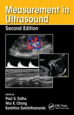
Measurement in Ultrasound
Seiten
2016
|
2nd edition
Crc Press Inc (Verlag)
978-1-4822-3135-9 (ISBN)
Crc Press Inc (Verlag)
978-1-4822-3135-9 (ISBN)
Measurement and interpretation of key ultrasound parameters are essential to differentiate normal anatomy from pathology. By using Measurement in Ultrasound, trainee radiologists and ultrasonographers can gain an appreciation of such measurements, while practitioners can use it as a valuable reference in the clinical setting.
The book follows a consistent format throughout for ease of reference and features useful information on preparation and positioning of the patient for ultrasound, the type of transducer and method to be used, the appearance of the resulting ultrasound images and the measurements to be derived from them.
Designed for frequent use in everyday practice, the book includes more than 150 high-quality ultrasound images annotated with key measurements and accompanied by concise explanatory text. Normal variants are provided, along with ranges for features that can change during development and in disease.
This new edition covers relevant developments in ultrasound. Where appropriate, updated ultrasound measurements that have arisen are also included and key references are provided as an aid to further study.
Edited by Paul S. Sidhu, BSc, MB BS, MRCP, FRCR, DTM&H FAIUM (Hon.), professor of imaging sciences and consultant radiologist, King’s College Hospital, London, UK Wui K. Chong, MB BS, MRCP, FRCR, assistant professor, Department of Radiology, University of North Carolina, Chapel Hill, USA Keshthra Satchithananda, BDS, FDSRCS, MB BS, FRCS, FRCR, consultant radiologist, King's College Hospital, London, UK
ADULTS. Upper Abdominal. Urinary Tract. Organ Transplantation. Genital Tract. Gastrointestinal Tract. Superficial Structures. Peripheral Vascular (Arterial). Peripheral Vascular (Venous). Musculoskeletal System. PEDIATRIC AND NEONATAL. Upper Abdomen. Renal Tract. Pediatric Uterus, Ovary, and Testis. Neonatal Brain. OBSTETRICS. Obstetrics.
| Zusatzinfo | 102 Tables, color; 185 Illustrations, color |
|---|---|
| Verlagsort | Bosa Roca |
| Sprache | englisch |
| Maße | 129 x 198 mm |
| Gewicht | 774 g |
| Themenwelt | Medizinische Fachgebiete ► Radiologie / Bildgebende Verfahren ► Radiologie |
| Medizinische Fachgebiete ► Radiologie / Bildgebende Verfahren ► Sonographie / Echokardiographie | |
| Studium ► 1. Studienabschnitt (Vorklinik) ► Anatomie / Neuroanatomie | |
| ISBN-10 | 1-4822-3135-2 / 1482231352 |
| ISBN-13 | 978-1-4822-3135-9 / 9781482231359 |
| Zustand | Neuware |
| Haben Sie eine Frage zum Produkt? |
Mehr entdecken
aus dem Bereich
aus dem Bereich
Buch (2023)
Thieme (Verlag)
190,00 €


