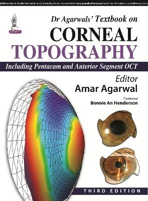
Dr Agarwal's Textbook on Corneal Topography
Jaypee Brothers Medical Publishers (Verlag)
978-93-5152-785-5 (ISBN)
Corneal topography is a non-invasive medical imaging technique for mapping the surface of the cornea. Dr Agarwal’s Textbook on Corneal Topography is the latest edition of this comprehensive guide to the capabilities of this type of imaging.
Divided into six sections, the first is an introduction to corneal topography and orbscan. The following sections cover specific imaging techniques and related issues including orbscan and refractive surgery, pentacam and anterior segment optical coherence tomography, aberropia, aberrations and topography, and refractive procedures and conditions. The final chapter covers corneal topography in cataract surgery.
This third edition is thoroughly updated with four brand new chapters. Section Three on pentacam and anterior segment OCT includes new chapters on corneal inflammation and optical coherence tomography, optical coherence tomography and corneal ectasia, and spectral-domain anterior segment optical coherence tomography in refractive surgery. The final section includes an OCT assessment of glued IOL position.
This highly illustrated guide to the latest developments in the field of corneal topography includes nearly 400 full colour images, making Dr Agarwal’s Textbook on Corneal Topography an essential resource for ophthalmologists.
Key Points
Comprehensive guide to the latest technological developments in corneal topography
New edition includes four brand new chapters
Nearly 400 full colour images and illustrations
Previous edition published 2010
Amar Agarwal Ms FRCS FRCOphth Chairman and Managing Director, Dr Agarwal’s Group of Eye Hospitals and Eye Research Center, Chennai, Tamil Nadu, India
Section I: Introduction to Corneal Topography and Orbscan
Fundamentals on Corneal Topography
Topographic Machines
Corneal Topography and the Orbscan
The Orbscan IIz Diagnostic System and Zywave Wavefront Analysis
Section II: Orbscan and Refractive Surgery
Orbscan Corneal Mapping in Refractive Surgery
Anterior Keratoconus
Posterior Corneal Changes in Refractive Surgery
Corneal Ectasia Post-LASIK: The Orbscan Advantages
Section III: Pentacam and Anterior Segment Optical Coherence Tomography
Pentacam
Evaluation of Patients for Refractive Surgery with Visante Anterior Segment OCT and the Combined Data Link with ATLAS Corneal Topographer
Corneal Inflammation and Optical Coherence Tomography
Optical Coherence Tomography in Corneal Ectasia
Spectral-domain Anterior Segment Optical Coherence Tomography in
Refractive Surgery
Section IV: Aberropia, Aberrations and Topography
Corneal Topographers and Wavefront Aberrometers: Complementary Tools
Aberrometry and Topography in the Vector Analysis of Refractive Laser Surgery
Aberropia: A New Refractive Entity
Differences between Various Aberrometer Systems
Corneal Wavefront Guided Excimer Laser Surgery for the Correction of Eye Aberrations
Ocular Higher Order Aberration Induced Decrease in Vision (Aberropia): Characteristics and Classification
Topographic and Aberrometer Guided Laser
NAVWave: Nidek Technique for Customized Ablation
Section V: Refractive Procedures and Conditions
Post-LASIK Latrogenic Ectasia
Decentered Ablations
Irregular Astigmatism: LASIK as a Correcting Tool
Posterior Chamber ICL and Toric ICL
Nidek OPD Scan in Clinical Practice
Section VI: Cataract
Corneal Topography in Cataract Surgery
Corneal Topography in Phakonit with a 5 mm Optic Rollable Intraocular Lens
Glued IOL Position: An OCT Assessment
Index
| Erscheint lt. Verlag | 10.5.2015 |
|---|---|
| Zusatzinfo | 367 Illustrations, unspecified; 17 Halftones, color |
| Verlagsort | New Delhi |
| Sprache | englisch |
| Maße | 216 x 279 mm |
| Gewicht | 950 g |
| Themenwelt | Medizin / Pharmazie ► Medizinische Fachgebiete ► Augenheilkunde |
| ISBN-10 | 93-5152-785-9 / 9351527859 |
| ISBN-13 | 978-93-5152-785-5 / 9789351527855 |
| Zustand | Neuware |
| Haben Sie eine Frage zum Produkt? |
aus dem Bereich


