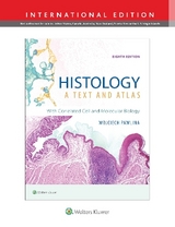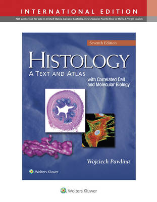
Histology: A Text and Atlas
With Correlated Cell and Molecular Biology
Seiten
2015
|
Seventh, International Edition
Lippincott Williams and Wilkins (Verlag)
978-1-4698-8931-3 (ISBN)
Lippincott Williams and Wilkins (Verlag)
978-1-4698-8931-3 (ISBN)
- Titel erscheint in neuer Auflage
- Artikel merken
Zu diesem Artikel existiert eine Nachauflage
Now in its seventh edition, Histology: A Text and Atlas is ideal for medical, dental, health professions, and undergraduate biology and cell biology students. This best-selling combination text and atlas includes a detailed textbook, which emphasizes clinical and functional correlates of histology fully supplemented by vividly informative illustrations and photomicrographs. Separate, superbly illustrated atlas sections follow almost every chapter and feature large-size, full-color digital photomicrographs with labels and accompanied descriptions that highlight structural and functional details of cells, tissues, and organs.
Updated throughout to reflect the latest advances in the field, this “two in one” text and atlas features an outstanding art program with all illustrations completely revised and redrawn as well as a reader-friendly format including red highlighted key terms, blue clinical text, and folders that cover clinical correlations and functional considerations.
NEW! All illustrations are now completely revised and redrawn for a consistent art program.
NEW! Histology 101 sections provide students with a reader-friendly review of essential information covered in the preceding chapters.
NEW! Updated cellular and molecular biology coverage reflects the latest advances in the field.
More than 100 atlas plates that incorporate 435 full-color, high-resolution photomicrographs.
Reader-friendly highlights including red bold terms, blue clinical text, and folders featuring clinical and functional correlations that increase student understanding and facilitates efficient study.
Easy-to-understand tables aid students in learning and reviewing information (such as staining techniques) without having to rely on rote memorization.
Features of cells, tissues, and organs and their functions and locations are presented in easy-to-locate, easy-to-review bulleted lists.
Additional clinical correlation and functional consideration folders have been added providing information related to symptoms, photomicrographs of diseased tissues or organs, short histopathological descriptions, and molecular basis for clinical intervention.
Methods
Cell Cytoplasm
The Cell Nucleus
Tissues: Concept and Classification
Epithelial Tissue
Connective Tissue
Cartilage
Bone
Adipose Tissue
Blood
Muscle Tissue
Nerve Tissue
Cardiovascular System
Lymphatic System
Integumentary System
Digestive System I: Oral Cavity and Associated Structures
Digestive System II: Esophagus and Gastrointestinal Tract
Digestive System III: Liver, Gallbladder, and Pancreas
Respiratory System
Urinary System
Endocrine Organs
Male Reproductive System
Female Reproductive System
Eye
Ear
| Erscheint lt. Verlag | 27.1.2015 |
|---|---|
| Zusatzinfo | 839 |
| Verlagsort | Philadelphia |
| Sprache | englisch |
| Maße | 213 x 276 mm |
| Gewicht | 2109 g |
| Themenwelt | Medizin / Pharmazie ► Gesundheitswesen |
| Medizin / Pharmazie ► Medizinische Fachgebiete | |
| Studium ► 1. Studienabschnitt (Vorklinik) ► Histologie / Embryologie | |
| ISBN-10 | 1-4698-8931-5 / 1469889315 |
| ISBN-13 | 978-1-4698-8931-3 / 9781469889313 |
| Zustand | Neuware |
| Haben Sie eine Frage zum Produkt? |
Mehr entdecken
aus dem Bereich
aus dem Bereich
Zytologie, Histologie und mikroskopische Anatomie
Buch | Hardcover (2022)
Urban & Fischer in Elsevier (Verlag)
54,00 €
Gewebelehre, Organlehre
Buch | Spiralbindung (2024)
Urban & Fischer in Elsevier (Verlag)
25,00 €
