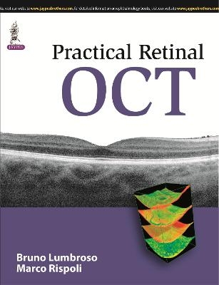
Practical Retinal OCT
Seiten
2014
Jaypee Brothers Medical Publishers (Verlag)
978-93-5152-532-5 (ISBN)
Jaypee Brothers Medical Publishers (Verlag)
978-93-5152-532-5 (ISBN)
Step by step guide to OCT for trainees. Includes chapters dedicated to 'en face' imaging and OCT for glaucoma, as well as qualitative and quantitative data analysis. Written by expert authors Bruno Lumbroso and Marco Rispoli.
Optical coherence tomography (OCT) is a non-invasive imaging test that uses light waves to take cross-section pictures of the retina, the light-sensitive tissue lining the back of the eye (eyeSmart).
This manual is a step by step guide to optical coherence tomography (OCT) imaging and its interpretation. Beginning with an overview of OCT and histology, the following chapter discusses three basic steps in normal OCT analysis – morphology study, retinal and choroidal structure study, and reflective study.
Qualitative and quantitative analysis of pathologic OCTs illustrates the evolution of various diseases, both spontaneous and after medical, surgical or laser therapy. Separate chapters are dedicated to ‘en face’ imaging and the role of OCT in the study and treatment of glaucoma.
Authored by renowned experts Bruno Lumbroso and Marco Rispoli from Centro Oftalmologico Mediterraneo in Rome, this practical reference is highly illustrated with clinical images, illustrations and tables throughout.
Key points
Step by step guide to OCT for trainees
Qualitative and quantitative data illustrates disease evolution, both spontaneous and after treatment
Separate chapters dedicated to ‘en face’ imaging and OCT for glaucoma
Written by expert ophthalmologists Bruno Lumbroso and Marco Rispoli
Bruno Lumbroso Marco Rispoli Both at Centro Oftalmologico Mediterraneo, Rome, Italy
Chapter 1: OCT and Histology
Chapter 2: Normal Chorioretinal OCT Analysis
Chapter 3: Qualitative Analysis of Pathologic OCTs
Chapter 4: Quantitative Analysis of Pathologic OCTs
Chapter 5: “En Face” Imaging
Chapter 6: Synthetic Study
Chapter 7: Reporting
Chapter 8: Glaucoma
| Erscheint lt. Verlag | 30.11.2014 |
|---|---|
| Zusatzinfo | 32 Tables, unspecified; 40 Halftones, unspecified; 45 Illustrations |
| Verlagsort | New Delhi |
| Sprache | englisch |
| Maße | 216 x 279 mm |
| Gewicht | 250 g |
| Themenwelt | Medizin / Pharmazie ► Medizinische Fachgebiete ► Augenheilkunde |
| ISBN-10 | 93-5152-532-5 / 9351525325 |
| ISBN-13 | 978-93-5152-532-5 / 9789351525325 |
| Zustand | Neuware |
| Haben Sie eine Frage zum Produkt? |
Mehr entdecken
aus dem Bereich
aus dem Bereich
Ein systematischer Ansatz
Buch | Hardcover (2022)
Urban & Fischer in Elsevier (Verlag)
265,00 €
1000 Fakten für die Facharztprüfung
Buch | Softcover (2023)
Springer (Verlag)
69,99 €


