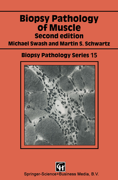
Biopsy Pathology of Muscle
Chapman and Hall (Verlag)
978-0-412-34880-8 (ISBN)
Introduction - general features of muscle, the motor unit, classifications of neuromuscular disorders, indications for muscle biopsy, selection for muscle biopsy, clinical features of neuromuscular disease, clinical investigation of neuromuscular disorders; the muscle biopsy - techniques and laboratory methods - preperation of the biopsy, cutting sections, histological methods, histological techniques for other structures found in muscle; histological and morphometric characteristics of normal muscle - fibre size, fibre-type distribution, fibre-type predominance, histological features; histological features of myopathic and neurogenic disorders - myopathic disorders, neurogenic disorders; inflammatory myopathies - clinical features of inflammatory myopathies, laboratory investigations, pathology, muscle involvement in other autoimmune disorders; muscular dystrophies - Duchenne muscular dysrophy, Becker muscular dystrophy, other X-linked dystrophies, limb-girdle muscular dystrophy, fascioscapulohumeral muscular dystrophy, Distal myopthies, myotonic dystrophy, Ocular myopathies and ocularpharyngeal dystrophy; 'benign' myopathies of childhood - Nemaline myopathy, central core disease, centronuclear (myotubular) myopathy, congenital fibre-type disproportion, myopathy with tubular aggregates, failure of fibre-types differentation, other benign myopathies of childhood, congenital muscular dystrophy; metabolic, endocrine and drug-induced myopathies - Metabolic myopathies, endocrine myopathies, drug-induced myopthies; neurogenic disorders - spinal muscular atropathies, motor neuron disease, other disorders of anterior horn cells and ventral roots, polyneuropathies, mononeuropathies; tumours of striated muscle and related disorders - primary tumours arising in muscle, tumours of muscle, fibrous tissue, fibrohistiocytic tumours, tumours of fat, blood vessels, peripheral nerves, other tumours that may arise in muscle, masses that mimic tumours (pseudotumours); interpretation of the muscle biopsy - is the biopsy abnormal?, myopathic or neurogenic?, specific morphological changes in myopthies, significance of some morphological abnormalities in muscle fibres, relation of pathological change to clinical disability or stage of disorder, are sequential biopsies useful?.
| Erscheint lt. Verlag | 4.9.1998 |
|---|---|
| Reihe/Serie | Biopsy Pathology Series |
| Zusatzinfo | 174 Illustrations, black and white; IX, 237 p. 174 illus. |
| Verlagsort | London |
| Sprache | englisch |
| Maße | 155 x 235 mm |
| Themenwelt | Medizin / Pharmazie ► Medizinische Fachgebiete ► Sportmedizin |
| Studium ► 1. Studienabschnitt (Vorklinik) ► Anatomie / Neuroanatomie | |
| Studium ► 2. Studienabschnitt (Klinik) ► Pathologie | |
| ISBN-10 | 0-412-34880-2 / 0412348802 |
| ISBN-13 | 978-0-412-34880-8 / 9780412348808 |
| Zustand | Neuware |
| Haben Sie eine Frage zum Produkt? |
aus dem Bereich


