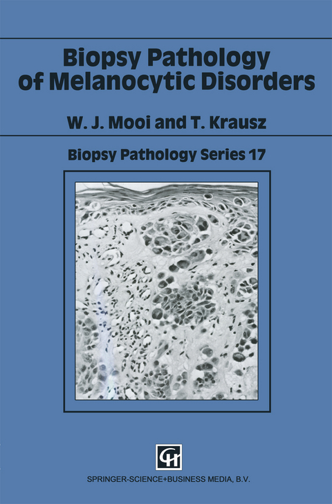
Biopsy Pathology of Melanocytic Disorders
Seiten
1998
|
Softcover reprint of the original 1st ed. 1992
Chapman and Hall (Verlag)
978-0-412-32350-8 (ISBN)
Chapman and Hall (Verlag)
978-0-412-32350-8 (ISBN)
Following sections on melanin and melanocytes and biopsy and histological investigation, this work discusses such topics as common acquired melanocytic naevi, congenital naevi, dysplastic naevus, cutaneous melanoma, prognostic factors and extracutaneous melanocytic lesions.
The incidence of cutaneous melanoma has risen considerably over the last few decades, and much attention is now paid to the early diagnosis of this tumour. As a consequence, pathologists are seeing more specimens of pigmented skin lesions. Clinically new entities, such as dysplastic naevi have been identified, and have added to the complexity of the differential diagnosis. Extracutaneous melanocytic lesions are less common but are of considerable clinical importance. This book is a further addition to the "Biopsy Pathology Series", and carries on the tradition of providing a well-illustrated guide for the reporting room pathologist. This has been achieved by highlighting those well-defined histological features which should be used as diagnostic criteria. In addition, tables of diagnostic features are also included, which should facilitate easy reference. This book should be of interest to pathologists and dermatologists.
The incidence of cutaneous melanoma has risen considerably over the last few decades, and much attention is now paid to the early diagnosis of this tumour. As a consequence, pathologists are seeing more specimens of pigmented skin lesions. Clinically new entities, such as dysplastic naevi have been identified, and have added to the complexity of the differential diagnosis. Extracutaneous melanocytic lesions are less common but are of considerable clinical importance. This book is a further addition to the "Biopsy Pathology Series", and carries on the tradition of providing a well-illustrated guide for the reporting room pathologist. This has been achieved by highlighting those well-defined histological features which should be used as diagnostic criteria. In addition, tables of diagnostic features are also included, which should facilitate easy reference. This book should be of interest to pathologists and dermatologists.
Melanin and melanocytes; biopsy, tissue processing and histological investigation; cutaneous pigmented lesions not related to melanocytic naevi; common acquired melanocytic naevi; cutaneous blue naevi and related lesions; congenital naevus; spitz naevus, desmoplastic spitz naevus and pigmented spindle cell naevus; dysplastic naevus; cutaneous melanoma; prognostic factors in cutaneous melanoma; extracutaneous melanocytic lesions; other extracutaneous melanotic lesions; cytological diagnosis of melanoma.
| Erscheint lt. Verlag | 4.9.1998 |
|---|---|
| Reihe/Serie | Biopsy Pathology Series |
| Zusatzinfo | 29 Illustrations, color; 254 Illustrations, black and white; XIII, 433 p. 283 illus., 29 illus. in color. |
| Verlagsort | London |
| Sprache | englisch |
| Maße | 155 x 235 mm |
| Themenwelt | Medizin / Pharmazie ► Medizinische Fachgebiete ► Dermatologie |
| Medizin / Pharmazie ► Medizinische Fachgebiete ► Onkologie | |
| Studium ► 2. Studienabschnitt (Klinik) ► Pathologie | |
| ISBN-10 | 0-412-32350-8 / 0412323508 |
| ISBN-13 | 978-0-412-32350-8 / 9780412323508 |
| Zustand | Neuware |
| Haben Sie eine Frage zum Produkt? |
Mehr entdecken
aus dem Bereich
aus dem Bereich


