Imaging Atlas of Human Anatomy
Seiten
2003
|
3rd Revised edition
Mosby (Verlag)
978-0-7234-3211-1 (ISBN)
Mosby (Verlag)
978-0-7234-3211-1 (ISBN)
- Titel erscheint in neuer Auflage
- Artikel merken
Zu diesem Artikel existiert eine Nachauflage
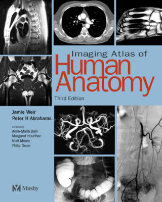
An atlas of normal anatomy as viewed through the range of imaging modalities. This third edition is organized by region, each area of normal anatomy being presented via a range of techniques. It includes development in imaging technology, particularly in terms of CT, MR and ultrasound imaging. It contains over 700 photographs, and 35 artworks.
Weir and Abrahams' "Imaging Atlas of Human Anatomy" is the definitive atlas of normal anatomy as viewed through the complete range of imaging modalities. The book is organized by region, each area of normal anatomy being presented via a range of techniques. The images are meticulously labeled, providing the reader with a reliable and comprehensive guide to normal human anatomy. The third edition has been updated to reflect advances in imaging technology, particularly in terms of CT, MR and ultrasound imaging. In all, 200 new diagnostic images have been added, and in response to user feedback, 25 new line diagrams have been added to aid interpretation of certain key images. The book therefore now includes over 700 photographs of outstanding clarity, as well as 35 interpretative artworks.
Weir and Abrahams' "Imaging Atlas of Human Anatomy" is the definitive atlas of normal anatomy as viewed through the complete range of imaging modalities. The book is organized by region, each area of normal anatomy being presented via a range of techniques. The images are meticulously labeled, providing the reader with a reliable and comprehensive guide to normal human anatomy. The third edition has been updated to reflect advances in imaging technology, particularly in terms of CT, MR and ultrasound imaging. In all, 200 new diagnostic images have been added, and in response to user feedback, 25 new line diagrams have been added to aid interpretation of certain key images. The book therefore now includes over 700 photographs of outstanding clarity, as well as 35 interpretative artworks.
Jamie Weir, MB BS DMRD FRCP (Ed) FRCR
Clinical professor of radiology at Aberdeen University, an enthusiastic teacher and writer, has written other titles for Mosby most notably Clinical Imaging (with Alison Murray).
Peter H. Abrahams , MB BS, FRCS (Ed), FRCR
Peter combines a part-time Family Practitioner job with academic positions at Girton College and Kigezi School in Cambridge, in addition to numerous publishing activities (ranging from Editorial Board of Clinical Anatomy to Consultant on Inside the Human Body). He has been involved in the publication of numerous Mosby anatomy titles, in print and non-print formats.
Introduction Head, neck, and brain Vertebral column and spinal cord Upper limb Thorax Abdomen Pelvis Lower limb
| Erscheint lt. Verlag | 2.7.2003 |
|---|---|
| Zusatzinfo | 760 ills. |
| Verlagsort | London |
| Sprache | englisch |
| Maße | 254 x 305 mm |
| Gewicht | 1115 g |
| Themenwelt | Medizinische Fachgebiete ► Radiologie / Bildgebende Verfahren ► Nuklearmedizin |
| Medizinische Fachgebiete ► Radiologie / Bildgebende Verfahren ► Radiologie | |
| Studium ► 1. Studienabschnitt (Vorklinik) ► Anatomie / Neuroanatomie | |
| ISBN-10 | 0-7234-3211-2 / 0723432112 |
| ISBN-13 | 978-0-7234-3211-1 / 9780723432111 |
| Zustand | Neuware |
| Haben Sie eine Frage zum Produkt? |
Mehr entdecken
aus dem Bereich
aus dem Bereich
Buch | Softcover (2022)
Lehmanns Media (Verlag)
39,95 €
Lehrbuch für Breast Care Nurses und Fachpersonen in der Onkologie
Buch | Hardcover (2020)
Hogrefe (Verlag)
50,00 €


