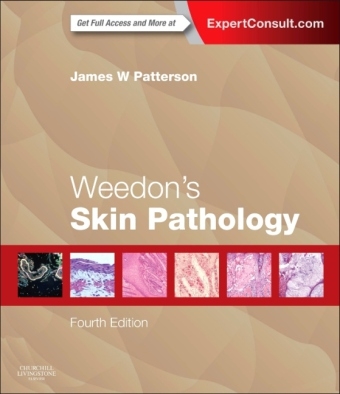
Weedon's Skin Pathology
Churchill Livingstone (Verlag)
978-0-7020-5183-8 (ISBN)
- Titel erscheint in neuer Auflage
- Artikel merken
New to this editon:
- Explore in-depth and updated topics covering clinically relevant developments in molecular biology and molecular techniques.
- Easily comprehend complex issues with improved illustrations focusing on rare conditions and unusual manifestations.
- Accurately interpret difficult specimens through an increased emphasis on differential diagnosis.
- Take advantage of expanded content in sections including Drug Reactions, Tumors, and Infections and Infestations.
- Expert Consult eBook version included with purchase. This enhanced eBook experience allows you to search all of the text, figures, references, and videos from the book on a variety of devices.
Key Features:
- Gain a full understanding of established disorders, unusual and rare disease entities, and incompletely defined entities.
- Immerse yourself in over 1,200 large-sized, high-quality illustrations.
- Provide the most accurate diagnoses possible with a design that reproduces what is seen through the microscope, thereby helping identify the characteristic features of the lesion demonstrated.
- Readily access important information through tables and boxes that organize diseases into groups, synthesize diagnostic criteria, and list differential diagnoses.
- Facilitate the identification of both key articles and more rare and unusual reports with remarkably authoritative, comprehensive, current, and relevant reference lists (over 35,000) for each entity.
By James W Patterson, MD, Professor of Pathology and Dermatology, Director of Dermatopathology, University of Virginia Health System,Charlottesville, VA
SECTION
1 INTRODUCTION
1 An approach to the interpretation of skin biopsies
2 Diagnostic clues SECTION
2 TISSUE REACTION PATTERNS
3 The lichenoid reaction pattern ('interface dermatitis!|)
4 The psoriasiform reaction pattern
5 The spongiotic reaction pattern
6 The vesiculobullous reaction pattern
7 The granulomatous reaction pattern
8 The vasculopathic reaction pattern SECTION
3 THE EPIDERMIS
9 Disorders of epidermal maturation and keratinization
10 Disorders of pigmentation SECTION
4 THE DERMIS
11 Disorders of collagen
12 Disorders of elastic tissue
13 Cutaneous mucinoses
14 Cutaneous deposits
15 Diseases of cutaneous appendages
16 Cysts, sinuses and pits
17 Panniculitis SECTION
5 THE SKIN IN SYSTE
MIC AN
D MISCELLANEOUS DISEASES
18 Metabolic and storage diseases
19 Miscellaneous conditions
20 Cutaneous drug reactions
21 Reactions to physical agents SECTION
6 INFECTIONS AN
D INFESTATIONS
22 Cutaneous infections and infestations -- histological patterns
23 Bacterial and rickettsial infections
24 Spirochetal infections
25 Mycoses and algal infections
26 Viral diseases
27 Protozoal infections
28 Marine injuries
29 Helminth infestations
30 Arthropod-induced diseases SECTION
7 TUMORS
31 Tumors of the epidermis
32 Lentigines, nevi and melanomas
33 Tumors of cutaneous appendages
34 Tumors and tumor-like proliferations of fibrous and related tissues
35 Tumors of fat
36 Tumors of muscle, cartilage and bone
37 Neural and neuroendocrine tumors
38 Vascular tumors
39 Cutaneous metastases
40 Cutaneous infiltrates -- non-lymphoid
41 Cutaneous infiltrates -- lymphoid and leukemic
| Zusatzinfo | Approx. 1250 illustrations (1246 in full color) |
|---|---|
| Verlagsort | London |
| Sprache | englisch |
| Gewicht | 4525 g |
| Einbandart | gebunden |
| Themenwelt | Medizin / Pharmazie ► Medizinische Fachgebiete ► Dermatologie |
| Studium ► 2. Studienabschnitt (Klinik) ► Pathologie | |
| ISBN-10 | 0-7020-5183-7 / 0702051837 |
| ISBN-13 | 978-0-7020-5183-8 / 9780702051838 |
| Zustand | Neuware |
| Informationen gemäß Produktsicherheitsverordnung (GPSR) | |
| Haben Sie eine Frage zum Produkt? |
aus dem Bereich


