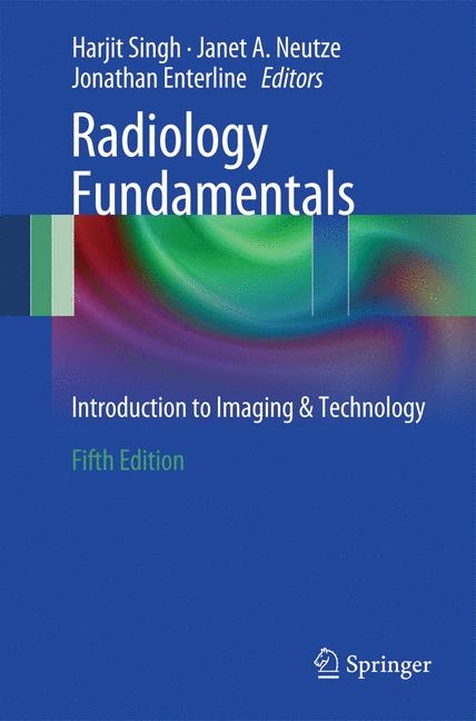
Radiology Fundamentals
Springer International Publishing (Verlag)
978-3-319-10361-7 (ISBN)
- Titel erscheint in neuer Auflage
- Artikel merken
Part 1. Introductory Section.- Chapter 1. Radiation Safety and Risk.- Chapter 2. Introduction to Radiology Concepts.- Chapter 3. Conventional Radiology.- Chapter 4. Ultrasound.- Chapter 5. Computed Tomography.- Chapter 6. MRI.- Chapter 7. Introduction to Nuclear Medicine.- Chapter 8. Cardiovascular and Interventional Radiology.- Part 2. Chest Section.- Chapter 9. Heart and Mediastinum.- Chapter 10. Coronary CTA.- Chapter 11. Lateral Chest.- Chapter 12. Pulmonary Mass Lesions.- Chapter 13. Air Space Disease.- Chapter 14. Interstitial Disease.- Chapter 15. Atelectasis.- Chapter 16. Pulmonary Vasculature.- Chapter 17. Pulmonary Edema.- Chapter 18. Pulmonary Embolism.- Chapter 19. Pneumothorax.- Chapter 20. Miscellaneous Chest.- Chapter 21. Tubes and Lines.- Part 3. Women's Section.- Chapter 22. Breast Imaging.- Chapter 23. Women’s Ultrasound.- Chapter 24. Women's Health Interventions.- Part 4. Abdominal Section.- Chapter 25. Abdominal Calficiations.- Chapter 26. Abdominal Harware and Tubes.- Chapter 27. Abnormal Air Collections of Abdomen.- Chapter 28. Bowel Obstruction.- Chapter 29. Concerning Abdominal Masses.- Chapter 30. Fluoroscopic Evaluation of the Upper GI Tract and Small Bowel.- Chapter 31. Imaging of the Colon.- Chapter 32. Imaging of the Gallbladder.- Chapter 33. Incidental Abdominal Lesion.- Chapter 34. Inflammatory and Infections Bowel Disease.- Chapter 35. Intra-abdominal Lymphadenopathy.- Part 5. Nuclear Medicine Section.- Chapter 36. Nuclear Medicine Cardiac Imaging.- Chapter 37. Gastrointestinal Nuclear Medicine.- Chapter 38. Oncological Nuclear Medicine.- Chapter 39. Pulmonary Nuclear Medicine.- Chapter 40. Skeletal Nuclear Medicine.- Part 6. Interventional Radiology Section.- Chapter 41. Diagnostic Arteriography.- Chapter 42. Pulmonary Arteriography and IVC Filter Placement.- Chapter 43. Percutaneous Nephrostomy Placement.- Chapter 44. TIPS.- Chapter 45. Central Venous Access.- Chapter 46. Interventional Oncology.- Part 7. Musculoskeletal Section.- Chapter 47. Fractures 1.- Chapter 48. Fractures 2.- Chapter 49. Bone Tumor Characteristics.- Chapter 50. Arthritis.- Part 8. Neuroradiology Section.- Chapter 51. CNS Anatomy.- Chapter 52. The Cervical Spine.- Chapter 53. Head Trauma.- Chapter 54. Stroke.- Chapter 55. Headache and Back Pain.- Part 9. Pediatrics Section.- Chapter 56. Pediatric Radiology Pearls.
"The purpose is to provide a general overview of the diagnostic imaging process while addressing common pathologies and diagnoses seen in patients. The audience is students who are entering the imaging profession. ... Overall, this is a useful book that provides a wide spectrum of information across the imaging sciences." (Kyle Theine, Doody's Book Reviews, April, 2015)
| Erscheint lt. Verlag | 15.12.2014 |
|---|---|
| Zusatzinfo | XVI, 374 p. 267 illus., 29 illus. in color. |
| Verlagsort | Cham |
| Sprache | englisch |
| Maße | 155 x 235 mm |
| Gewicht | 697 g |
| Themenwelt | Medizinische Fachgebiete ► Radiologie / Bildgebende Verfahren ► Nuklearmedizin |
| Medizinische Fachgebiete ► Radiologie / Bildgebende Verfahren ► Radiologie | |
| Schlagworte | computed tomorgraphy • introduction to radiology • Magnetic Resonance Imaging • medical use of imaging modalities • Nuclear Medicine • Radiologie • role of radiology in diagnosis and treatment |
| ISBN-10 | 3-319-10361-X / 331910361X |
| ISBN-13 | 978-3-319-10361-7 / 9783319103617 |
| Zustand | Neuware |
| Haben Sie eine Frage zum Produkt? |
aus dem Bereich


