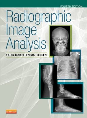
Radiographic Image Analysis
Seiten
2014
|
4th edition
Saunders (Verlag)
978-0-323-28052-5 (ISBN)
Saunders (Verlag)
978-0-323-28052-5 (ISBN)
- Titel erscheint in neuer Auflage
- Artikel merken
Zu diesem Artikel existiert eine Nachauflage
This comprehensive guide provides all the tools you need to accurately evaluate radiographic images and make the adjustments needed to acquire the best possible diagnostic quality images. Youll discover how to evaluate an image, identify any improper positioning or techniques that caused poor quality, and correct the problem. No other text is devoted to equipping you with the critical thinking skills needed to properly position patients for optimal radiographs and help minimize the need for repeat images.
"The whole text is well presented." Reviewed by Jenny May on behalf of Radiography, July 2015
Poorly positioned example images appear at the end of procedures to test your knowledge.
Spotlights concepts boxes highlight the most important information as it appears in the chapters and directs readers to more information on these topics.
Chapter objectives, key terms, and outlines help in mastering important concepts and information.
NEW! Expanded sections on pediatric, obesity, and trauma digital radiography provides the most pertinent and up-to-date information needed for clinical success.
NEW! Reformatted content surrounding procedures includes the following to help you identify correctly and incorrectly positioned patients:
accurately positioned projection with labeled anatomy
photograph of an accurately positioned model
table that provides a detailed one-to-one correlation between the positioning procedures and image analysis guidelines
discussion, with correlating images, on identifying how the patient, central ray, or image receptor were poorly positioned if the projection does not demonstrate an image analysis guideline
discussion of topics relating to positioning for patient condition variations and non-routine situations
photographs of bones and models positioned as indicated to clarify information and demonstrate anatomy alignment when distortion makes it difficult
practice images of the projection that demonstrate common procedural errors
NEW! Two-color design helps you read and retain pertinent information.
NEW! Updated boxed material summarizes important analysis details and provides a quick reference.
NEW! Highlighted table data offers a new format to aid in the understanding of field size requirements using direct-capture digital radiography.
1. Image Analysis Guidelines
2. Digital Imaging Guidelines
3. Image Analysis of the Chest and Abdomen
4. Image Analysis of the Upper Extremity
5. Image Analysis of the Shoulder
6. Image Analysis of the Lower Extremity
7. Image Analysis of the Pelvis, Hip, and Sacroiliac Joints
8. Image Analysis of the Cervical and Thoracic Vertebrae
9. Image Analysis of the Lumbar Vertebrae, Sacrum, and Coccyx
10. Image Analysis of the Sternum and Ribs
11. Image Analysis of the Cranium, Facial Bones, Paranasal Sinuses
12. Image Analysis of the Digestive System
Bibliography
Glossary
| Erscheint lt. Verlag | 27.1.2015 |
|---|---|
| Zusatzinfo | Approx. 1215 illustrations; Illustrations |
| Verlagsort | Philadelphia |
| Sprache | englisch |
| Maße | 216 x 276 mm |
| Gewicht | 1700 g |
| Themenwelt | Medizin / Pharmazie ► Gesundheitsfachberufe ► MTA - Radiologie |
| ISBN-10 | 0-323-28052-8 / 0323280528 |
| ISBN-13 | 978-0-323-28052-5 / 9780323280525 |
| Zustand | Neuware |
| Haben Sie eine Frage zum Produkt? |
Mehr entdecken
aus dem Bereich
aus dem Bereich
Buch | Softcover (2024)
Studia Universitätsverlag Innsbruck
48,00 €
Buch | Hardcover (2024)
Mosby (Verlag)
229,40 €


