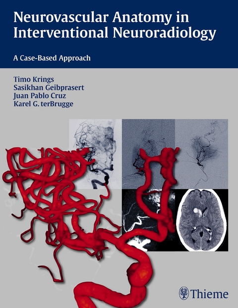
Neurovascular Anatomy in Interventional Neuroradiology
Thieme Medical Publishers Inc (Verlag)
978-1-60406-839-9 (ISBN)
The case discussions include modern examples of invasive and non- invasive angiographic techniques that are relevant for general radiologists and diagnostic neuroradiologists as well as interventionalists. This book gives readers the detailed knowledge of neurovascular anatomy that allows them to anticipate and avoid potential complications.
Key Features:
- Cases are enhanced by more than 1,000 high-quality radiographs covering the full range of neurovascular anatomy
- Content focuses on the practical relevance of the anatomical features encountered while performing everyday neurovascular procedures
- Anatomy and embryology are explained together, enabling readers to fully comprehend the vascular anatomy and its many variants
- Pearls and pitfalls are provided at the end of each chapter, highlighting the critical anatomy points presented
All neuroradiologists, interventionalists, general radiologists, and diagnostic neuroradiologists, as well as residents and fellows in these specialties, will read this book cover to cover and frequently consult it for a quick review before performing procedures.
Associate Professor, Division of Neuroradiology, University of Toronto, Toronto, Ontario, Canada University of Toronto, Toronto, Ontario, Canada Hospital for Sick Children and Toronto Western Hospital, University of Toronto, Toronto, ON
Section I: Aortic Arch
Case 1: The Common Origin of the Brachiocephalic and Left Common Carotid Artery
Case 2: The Aberrant Subclavian Artery
Section II: Internal Carotid Artery
Case 3: The Carotid Segments, the Aberrant ICA, and the Persistent Stapedial Artery
Case 4: Persistent Carotid-Vertebrobasilar Anastomoses
Case 5: The Inferolateral and the Meningohypophyseal Trunk
Case 6: The Dural Ring and the Carotid Cave
Case 7: The Dorsal and Ventral Ophthalmic Arteries
Case 8: The Branches of the Ophthalmic Artery
Case 9: The Anterior Choroidal Artery
Section III: Anterior Circulation
Case 10: The Infraoptic Course of the Anterior Cerebral Artery
Case 11: The Anterior Communicating Artery Complex
Case 12: The Azygos Anterior Cerebral Artery
Case 13: The Cortical Branches of the Anterior Cerebral Artery
Case 14: The Middle Cerebral Artery Trunk
Case 15: The Recurrent Artery of Heubner
Case 16: The Cortical Branches of the Middle Cerebral Artery
Case 17: The Leptomeningeal Anastomoses
Section IV: Posterior Circulation
Case 18: Variations of the Origin of the PICA
Case 19: The Cerebellar Arteries
Case 20: The Basilar Artery Trunk
Case 21: The Brainstem Perforators
Case 22: The Basilar Tip
Case 23: The Thalamoperforating Arteries
Case 24: The Cortical Branches of the Posterior Cerebral Artery
Section V: External Carotid Artery
Case 25: The "Dangerous" Anastomoses I: Ophthalmic Anastomoses
Case 26: The "Dangerous" Anastomoses II: Petrous and Cavernous Anastomoses
Case 27: The "Dangerous" Anastomoses III: Upper Cervical Anastomoses
Case 28: The Cranial Nerve Supply
Case 29: The Vascular Anatomy of the Nose
Case 30: The Ascending Pharyngeal Artery
Case 31: The Meningeal Supply
Section VI: Cerebral Veins
Case 32: The Superior Sagittal and Transverse Sinuses
Case 33: The Cavernous Sinus
Case 34: The Superficial Cortical Veins
Case 35: The Transmedullary Veins
Case 36: The Deep Venous System I: Internal Cerebral Veins, Tributaries, and Drainage
Case 37: The Deep Venous System II: The Basal Vein of Rosenthal and the Venous Circle
Case 38: The Infratentorial Veins
Section VII: Spine
Case 39: The Segmental Spinal Arteries
Case 40: The Radiculopial and Radiculomedullary Arteries
Case 41: The Intrinsic Arteries of the Cord
Case 42: The Artery of the Filum Terminale
Case 43: The Spinal Cord Veins
| Zusatzinfo | 1295 Abbildungen |
|---|---|
| Verlagsort | New York |
| Sprache | englisch |
| Gewicht | 834 g |
| Einbandart | kartoniert |
| Themenwelt | Medizinische Fachgebiete ► Chirurgie ► Neurochirurgie |
| Medizin / Pharmazie ► Medizinische Fachgebiete ► Neurologie | |
| Medizinische Fachgebiete ► Radiologie / Bildgebende Verfahren ► Nuklearmedizin | |
| Medizinische Fachgebiete ► Radiologie / Bildgebende Verfahren ► Radiologie | |
| Schlagworte | diagnostische Neuroradiologie • Interventionelle Untersuchungen • invasive Röntgendiagnostik • klinische Neuroanatomie • Neuroanatomie • Neuroradiologie • Neurovascular Imaging • Neurovaskuläre Krankheiten • Radiologie: Interventionelle Radiologie |
| ISBN-10 | 1-60406-839-6 / 1604068396 |
| ISBN-13 | 978-1-60406-839-9 / 9781604068399 |
| Zustand | Neuware |
| Informationen gemäß Produktsicherheitsverordnung (GPSR) | |
| Haben Sie eine Frage zum Produkt? |
aus dem Bereich


