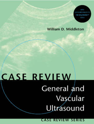
General and Vascular Ultrasound
Mosby (Verlag)
978-0-323-00736-8 (ISBN)
- Titel erscheint in neuer Auflage
- Artikel merken
This title is perfect as a study tool or a quick reference! This new resource presents realistic case studies in a unique question-and-answer format to help assess readers' knowledge in this sub speciality, pass certification exams, and succeed in practice. It's practical organization and outstanding illustrations make it easy to hone clinical skills.
I.Opening Round E1: Normal Kidneys E2: Gallbladder Sludge E3: Angiomyolipoma E4: Normal Anatomy of the Liver E5: Normal Anatomy of the Common Bile Duct E6: Pleural Effusion E7: Normal Scrotal Anatomy E8: Normal Peripancreatic Anatomy E9: Hydronephosis E10:Normal Liver and Gallbladder Anatomy E11:Hepatic Target Lesions E12:Normal Anatomy of the Shoulder E13:Gallbladder Wall Thickening E14:Normal Hepatic Venous Anatomy E15:Variant Relationship of Right Hepatic Artery and Bile Duct E16:Normal Anatomy of the Thyroid E17:Normal Prostate E18:Complete Tear of the Achilles Tendon E19:Orchitis E20:Transitional Cell Carcinoma of the Bladder E21:Splenomegaly E22:Thyroglossal Duct Cyst E23:High Resistance Waveforms E24:Ganglion Cyst of the Wrist E25:Acute Appendicitis E26:Distance Gain Compensation Curve E27:Abdominal Aortic Aneurysm E28:Liver Metastases E29:Ascites E30:Renal Stones E31:Testicular Cysts E32:Doppler Aliasing Artifact E33:Foreign Body E34:Nodular Hyperplasia of the Thyroid E35:Junctional Parenchymal Defect E36:Adenocarcinoma of the Pancreas E37:Epididymitis E38:Distal Uretal Stone E39:Lower Extremity Deep Vein Thrombosis E40:Normal Carotid Bifurcation E41:Testicular Seminoma E42:Autosomal Dominant Polycystic Kidney Disease E43:Acute Cholecystitis E44:Mixed Germ-Cell Tumors E45:Extrahepatic Billlary Dilation E46:Renal Cysts E47:Pancreatic Pseudocysts E48:Frame Rate and Resolution E49:Hepatic Hemangioma E50:Ruptured Bakers Cyst E51:Intrahepatic Biliary Ductal Dilation E52:Hydrocele E53:Parathyroid Adenoma E54:Choledocholithiasis E55:Fatty Infiltration of the Liver E56:Techinical Parameters Important in Producing Shadowing From Small Gallstones E57:Renal Parenchymal Disease E58:Spermatocele E59:Acute Pancreatitis E60:Tumefactove Sludge (Sludgeball) E61:Varicocele E62:Beam Steering and Color Assignment E63:Post Catheterization Pseudoaneurysm E64:Splenic Infiltration E65:Replaced Right Hepatic Artery E66:Floating Gallstones II.Fair Game M1: Porcelain Gallbladder M2: Calcific Tendonitis of the Rotator Cuff M3: Adenomatous Polyps of the Gallbladder M4: Cholesterol Polyps of the Gallbladder M5: Mirror Image Artifact M6: Gallbladder Cancer M7: High Grade Carotid Stenosis M8: Comparison of Color Doppler and Power Doppler M9: Rectus Sheath Hematoma M10:Comparison of Phased Array and Curved Array Transducers M11:Normal Relationship of Pancreatic Tail and Spleen M12:Focal Splenic Lesions M13:Testicular Torsion M14:Subclavian Steal M15:Mortons Neuroma M16:Partially Calcified Liver Metastases M17:Renal Transplant Lymphocele M18:Doppler Mirror Image Artifact M19:Tenosynovitis M20:Papillary Thyroid Cancer M21:Inguinal Adenopathy M22:Normal Variant Pancreatic Head M23:Tunica Albuginea Cyst M24:Renal Cell Carcinoma M25:Cirrhosis M26:Subclavian Vein Obstruction M27:Umbilical Vein Collateral M28:Splenule M29:Ectopic Parathyroid Adenoma M30:Prostate Cancer M31:Low-Grade Internal Carotid Stenosis M32:Effect of Power Output on Color Doppler Images M33: Hepatic Subcapsular Hematoma M34:Normal Tendon Anisotropy M35:Intrabiliary Air M36:Plantar Fasciitis M37:Tumor Thrombus of the Renal Vein and IVC M38:Effect of Doppler Scale on Doppler Sensitivity M39:Tissue Vibration M40:Wall-Echo-Shadow Complex M41:Hepatic Abscess M42:Portal Vein Thrombosis M43:Lymphoma M44:Stones Impacted in the Gallbladder Neck M45:Medullary Nephrocalcinosis M46:Normal Hepatic Venous Waveform M47:Mucinos Macrocystac Pancreatic Neoplasm M48:Testicular Abscess M49:Replaced Left Hepatic Artery M50:Aliasing M51:Portal Vein Flow Reversal M52:Intrahepatic Bile Duct Stones M53:Intusssception M54:Prostatic Cyst M55:Leydig Cell Tumor M56:Undescended Testis M57:Renal Abscess M58:Changes in Color Shading M59:Use of the Wall Filter to Suppress Tissue Motion M60:Islet Cell Tumor of the Pancreas M61:Spectral Doppler Measurements M62:Full Thickness Rotator Cuff Tear M63:Focal Fatty Infiltration of the Liver M64:Femoral Arterial Venous Fistula M65:Ring-down Artifact M66:Complex Hydroceles M67:Classic Testicular Microlithiasis M68:Multilocular Cystic Nephroma M69:Low Resistance Arterial Waveforms With and Without Spectral Broadening M70:Fatty Infiltration with Focal Spring M71:Chronic Pancreatitis M72:Tissue Harmonic Imaging M73:Relationship of Velocity Calculation Accuracy to Doppler Angle M74:Tardus Parvus Waveform M75:Cholangiocarcinoma M76:Gallstones III.Challenge H1: Hepatitis H2: Splenic Sarcoid H3: Hepatic Focal Nodular Hyperplasia H4: Hashimotos Thyroiditis H5: Adrenal Masses H6: Peyronnies Disease H7: Full Thickness Rotator Cuff Tear H8: Transitional Cell Carcinoma H9: Liver Laceration and Hemoperitoneum H10:Peritoneal Metastases H11:Normal Testicular Vasculature H12:Complete Occlusion of the Internal Carotid H13:Von Hippel-Lindau Disease H14:Bile Duct Wall Thickening H15:Intraoperative Scans of Pancreatic Islet Cell Tumors H16:Side Love Artifact H17:Carolis Disease H18:Normal Variant: Left Hepatic Lobe Over Spleen H19:Gastric Wall Thickening H20:Giant Cell Tumor of the Tendon Sheath H21:Gangrenous Cholecystitis H22:Tubular Ectasia of the Rete Testes H23:Increased Renal Artery Velocity Due to Arterial Stenosis H24:Common Carotid Occlusion H25:Post Traumatic Renal Pseudoaneurysm and Arteriovenous Fistula H26:Echinococcal Cyst H27:Splenosis H28:Effect of Color Priority on Color Doppler Images H29:Reversed Flow in the Coronary Vein H30:Horseshoe Kidney H31:Epidermoid Cyst H32:Renal Vein Thrombosis H33:Effect of Transmit Frequency on Doppler Sensitivity H34:Partial Thickness Rotator Cuff Tear H35:Subacute Thyroiditis H36:Page Kidney H37:Midline Refraction Artifact H38:Blunted Intrarenal Artery Waveform Due to Renal Artery Stenosis H39:Biceps Tendon Dislocation H40:Effect of Beam Steering on Doppler Sensitivity H41:Helical Flow H42:Renal Vascular Variants H43:Partial Longitudinal Tendon Tear H44:Adenomyomatosis of the Gallbladder H45:Peripelvic Cysts Simulating Hydronephrosis H46:Chronic DVT H47:Tumor Thrombus in the Portal Vein H48:Acquired Cystic Disease H49:Measurement of Flow Volume H50:Emphysematous Cholecystitis H51:Biceps Tendon Rupture H52:Effect of Frame Rate on Color Doppler Images H53:Cavernous Transformation of the Portal Vein H54:Vascular Invasion from Pancreatic Cancer H55:Prominent Papillary Tips H56:Diaphragmatic Duplication Artifact H57:Muscle Tear and Hematoma H58:Hepatic Artery Thrombosis Status Post Liver Transplant H59:Ulcerated Carotid Plaque H60:Urinary Obstruction H61:Passive Hepatic Congestion H62:Microcystic Serous Cystadenoma of the Pancreas H63:Hepatic Vein Thrombosis H64:Fibromuscular Dysplasia H65:Pyelmonephritis H66:Complex Renal Cysts H67:Urethral Diverticulum H68:Stenosis of a TIPS Stent
| Erscheint lt. Verlag | 6.11.2001 |
|---|---|
| Reihe/Serie | Case Review |
| Zusatzinfo | 483 ills |
| Verlagsort | London |
| Sprache | englisch |
| Maße | 216 x 279 mm |
| Gewicht | 788 g |
| Themenwelt | Medizinische Fachgebiete ► Radiologie / Bildgebende Verfahren ► Sonographie / Echokardiographie |
| ISBN-10 | 0-323-00736-8 / 0323007368 |
| ISBN-13 | 978-0-323-00736-8 / 9780323007368 |
| Zustand | Neuware |
| Haben Sie eine Frage zum Produkt? |
aus dem Bereich


