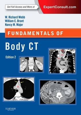
Fundamentals of Body CT
Seiten
2014
|
4th Revised edition
Saunders (Verlag)
978-0-323-22146-7 (ISBN)
Saunders (Verlag)
978-0-323-22146-7 (ISBN)
- Titel ist leider vergriffen;
keine Neuauflage - Artikel merken
Zu diesem Artikel existiert eine Nachauflage
Perfect for radiology residents and practitioners, Fundamentals of Body CT offers an easily accessible introduction to body CT! Completely revised and meticulously updated, this latest edition covers today's most essential CT know-how, including the use of multislice CT to diagnose chest, abdominal, and musculoskeletal abnormalities, as well as the expanded role of 3D CT and CT angiography in clinical practice. It's everything you need to effectively perform and interpret CT scans.
"I would recommend this book to junior doctors/AHPs with an interest in CT and its applications, and to those diagnostic consultants who report body CT while specialising in other areas." Reviewed by RAD Magazine, Apr 2015
"..all aspects of this book are relevant to CT practitioners and it is potentially a useful source for students." Reviewed by Louise Mifsud on behalf of Radiography Journal, April 2015
Glean all essential, up-to-date, need-to-know information to effectively interpret CTs and the salient points needed to make accurate diagnoses.
Review how the anatomy of each body area appears on a CT scan.
Grasp each procedure and review key steps quickly with a comprehensive yet concise format.
Achieve optimal results with step-by-step instructions on how to perform all current CT techniques.
Compare diagnoses with a survey of major CT findings for a variety of common diseases-with an emphasis on those findings that help to differentiate one condition from another.
Make effective use of 64-slice MDCT and dual source CT scanners with coverage of the most current indications.
Stay current extensive updates of clinical guidelines that reflect recent changes in the practice of CT imaging, including (ACCP) Diagnosis and Management of Lung Cancer guidelines, paraneoplastic and superior vena cava syndrome, reactions to contrast solution and CT-guided needle biopsy.
Get a clear view of the current state of imaging from extensively updated, high-quality images throughout.
Access the complete contents online, fully searchable, at ExpertConsult.
Part I: Thorax
1. Intro to CT of the Thorax: Chest CT Techniques
2. Mediastinum-Introduction & Normal Anatomy
3. Mediastinum-Vascular Abnormalities
4. Mediastinum- Lymph Node Abnormalities & Masses
5. The Pulmonary Hila
6. Lung Disease
7. Pleura, Chest Wall, and Diaphragm
Part II: The Abdomen & Pelvis
8. Introduction to CT of the Abdomen & Pelvis
9. Peritoneal Cavity, Vessels, Nodes, and Abdominal Wall
10. Abdominal Trauma
11. Liver
12. Biliary Tree and Gallbladder
13. Pancreas
14. Spleen
15. Kidneys and Ureters
16. Adrenal Glands
17. Gastrointestinal Tract
18. Pelvis
Part III: Musculoskeletal Skeleton
19. CT in Musculoskeletal Trauma
20. CT in Musculoskeletal Nontrauma
21. Incidental Findings
| Reihe/Serie | Fundamentals of Radiology |
|---|---|
| Zusatzinfo | Approx. 350 illustrations |
| Verlagsort | Philadelphia |
| Sprache | englisch |
| Maße | 184 x 260 mm |
| Themenwelt | Medizinische Fachgebiete ► Radiologie / Bildgebende Verfahren ► Computertomographie |
| Medizinische Fachgebiete ► Radiologie / Bildgebende Verfahren ► Radiologie | |
| ISBN-10 | 0-323-22146-7 / 0323221467 |
| ISBN-13 | 978-0-323-22146-7 / 9780323221467 |
| Zustand | Neuware |
| Haben Sie eine Frage zum Produkt? |
Mehr entdecken
aus dem Bereich
aus dem Bereich


