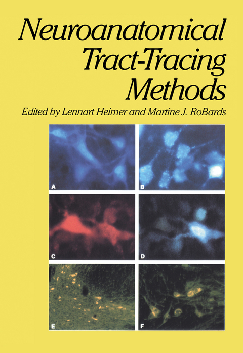
Neuroanatomical Tract-Tracing Methods
Springer-Verlag New York Inc.
978-1-4613-3191-9 (ISBN)
, 1962) developed a fluorescent histochemical method to visualize mono- amine-containing cells in the brain; this technique was soon applied to show that the rich dopaminergic terminal field in the striatum derived from neu- rons in the substantia nigra (Anden et at. , 1964). In the following decade, refinements in the histofluorescent method and the development of sensitive silver impregnation methods permitted a detailed light microscopic explo- ration of the dopaminergic nigrostriatal system.
1 Experimental Neuroanatomy: General Approaches and Laboratory Procedures.- I. Introduction.- II. Tract-Tracing Methods.- III. Practical Problems.- IV. Analysis.- V. The Neuroanatomical Laboratory.- VI. Appendix.- 2 Methods for Selective, Restricted Lesion Placement in the Central Nervous System.- I. Introduction.- II. Stereotaxic Technique.- III. Nonselective Lesion Techniques.- IV. Evaluation of the Electrolytic Lesion.- V. Selective Lesion Techniques.- VI. The Interpretation of Lesion Effects.- VII. Appendix: Stereotaxic Atlases.- 3 Methods for Delivering Tracers.- I. Introduction.- II. Pressure Injection.- III. Iontophoretic Injection.- IV. Appendix.- 4 Silver Methods for the Impregnation of Degenerating Axoplasm.- I. Introduction.- II. Theoretical Considerations.- III. Practical Aspects.- IV. General Characteristics of the Silver Methods.- V. Interpretation of Degenerating Fibers and Terminal Degeneration.- VI. Other Degenerative Neuronal Phenomena.- VII. Sources of Error.- VIII. Summary of Advantages and Limitations.- IX. Prospects for the Future.- X. Appendix.- 5 The Autoradiographic Tracing of Axonal Connections in the Central Nervous System.- I. Introduction.- II. The Principles of the Method.- III. Methodology.- IV. Analysis of the Data.- V. Electron Microscopic Autoradiography.- VI. Summary of Advantages and Limitations.- VII. Appendix.- 6 Horseradish Peroxidase: The Basic Procedure.- I. Introduction.- II. Basic Applications.- III. Incorporation and Transport of HRP.- IV. Methodology.- V. General Characteristics of the Different HRP Methods.- VI. Results and Interpretations.- VII. Summary of Advantages and Limitations.- VIII. Appendix.- 7 Horseradish Peroxidase: Intracellular Staining of Neurons.- I. Introduction.- II. Methods.- III. Application of the Technique.- IV. Summary of Advantages and Limitations.- V. Appendix.- 8 Horseradish Peroxidase and Fluorescent Substances and Their Combination with Other Techniques.- I. Introduction.- II. The Tracing of Collateral Projections.- III. HRP and Anterograde Tracing Methods.- IV. HRP and Transmitter-Related Histochemical Procedures.- V. HRP and 2-Deoxyglucose Procedures.- VI. Prospects for the Future.- VII. Appendix.- 9 The Golgi Methods.- I. Introduction.- II. The Rapid Golgi Method.- III. Analysis of the Data.- IV. Presentation of the Data.- V. Variations of the Golgi Method.- VI. Summary of Advantages and Limitations.- VII. Appendix.- 10 Electron Microscopy: Preparation of Neural Tissues for Electron Microscopy.- I. Introduction.- II Basic Procedures for Fixation and Embedding.- III. Variations.- IV. Evaluation of Results with the Light Microscope.- V. Cutting and Staining Ultrathin Sections.- VI. Synthesis.- VIII. Appendix.- 11 Electron Microscopy: Identification and Study of Normal and Degenerating Neural Elements by Electron Microscopy.- I. Introduction.- II. Bridging the Gap between Light and Electron Microscopy.- III. Practical Guidelines for Electron Microscopy.- IV. Identification of Neuronal Elements.- V. Ultrastructure of Degenerating Nerve Fibers.- VI. Morphometry.- 12 Tract Tracing by Electron Microscopy of Golgi Preparations.- I. Introduction.- II. General Description of Techniques.- III. Summary of Advantages and Limitations.- IV. Concluding Comments and Troubleshooting.- V. Appendix.- 13 Fluorescence Histochemical Methods: Neurotransmitter Histochemistry.- I. Introduction.- II Chemical Basis of the Fluorescence Histochemical Methods.- III. Equipment.- IV. Methods Using Formaldehyde or Glyoxylic Acid Condensation.- V. The Selection of Fluorescence Histochemical Methods.- VI. Advantages and Limitations of the Fluorescence Histochemical Method.- VII. Appendix.- 14 Immunocytochemical Methods.- I. Introduction.- II. Types of Immunocytochemical Techniques.- III. The Peroxidase-Antiperoxidase (PAP) Technique.- IV. Variations in PAP Technique.- V. Specificity of the PAP Technique.- VI. Use of the PAP Technique in the Demonstration of Catecholaminergic Neurons.- VII. Use of the PAP Technique in the Localization of Neuropeptides.- VIII Summary of Advantages and Limitations of the PAP Technique.- IX. Conclusions.- 15 The 2-Deoxyglucose Method.- I. Introduction.- II. Basic Principles of the Method.- III. General Applications of the Method.- IV. Methodology for [14C]-2DG.- V. Methodology for [3H]-2DG.- VI. Data Analysis.- VII. Advantages and Limitations.- VIII Appendix.- Epilogue: Some General Advice to the Young Investigator.- Author Index.
| Zusatzinfo | XXIII, 567 p. |
|---|---|
| Verlagsort | New York, NY |
| Sprache | englisch |
| Maße | 170 x 244 mm |
| Themenwelt | Medizin / Pharmazie ► Studium |
| Naturwissenschaften ► Biologie ► Humanbiologie | |
| Naturwissenschaften ► Biologie ► Zoologie | |
| ISBN-10 | 1-4613-3191-9 / 1461331919 |
| ISBN-13 | 978-1-4613-3191-9 / 9781461331919 |
| Zustand | Neuware |
| Haben Sie eine Frage zum Produkt? |
aus dem Bereich


