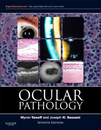
Ocular Pathology
W B Saunders Co Ltd (Verlag)
978-1-4557-2874-9 (ISBN)
- Titel erscheint in neuer Auflage
- Artikel merken
"This seventh edition of Ocular Pathology by Myron Yanoff and Joseph Sassani is a superb update of what has become the single best ophthalmic pathology reference text for ophthalmologists, pathologists and researchers." Foreword by: J. Douglas Cameron, Ophthalmology and Visual Neurosciences, University of Minnesota School of Medicine,�June 2015
Take advantage of clinical "pearls" that offer you the benefits of proven strategies.
Quickly reference information with help from a convenient outline format, ideal for today's busy physician.
Visualize every concept by viewing 1,900 illustrations, 1,600 of which are in full color, from the collections of internationally renowned leaders in ocular pathology.
Understand the role of VEGF and other factors in the pathobiology of diabetic complications, as well as the pathobiology of myocilin and the TIGR gene in the development of glaucoma.
Review the latest features related to the pathobiology of central corneal thickness.
Stay abreast of the latest in ocular pathology with coverage of the classification system for retinoblastoma; immunopathology of herpes keratitis; and genetic features of persistent hyperplastic primary vitreous.
Access the entire text online at Expert Consult, and test your visual recognition and understanding of disease with a new online image review/testing feature.
1. BASIC PRINCIPLES OF PATHOLOGY
Inflammation
Immunobiology
Cellular and Tissue Reactions
2. CONGENITAL ANOMALIES
Phakamatoses (Disseminated Hereditary Hamartomas)
Chromosomal Aberrations
Infectious Embryopathy
Drug Embryopathy
Other Congenital Anomalies
3. NONGRANULOMATOUS INFLAMMATION:
UVEITIS, ENDOPHTHALMITIS, PANOPHTHALMITIS, AND SEQUELAE
Classification
Suppurative Endophthalmitis and Panophthalmitis
Nonsuppurative, Chronic Nongranulomatous Uveitis and Endophthalmitis
Sequelae of Uveitis, Endophthalmitis, and Panophthalmitis
End Stage of Diffuse Ocular Diseases
4. GRANULOMATOUS INFLAMMATION
Introduction
Post-Traumatic
Nontraumatic Infectious
Nontraumatic Noninfectious
5. SURGICAL AND NONSURGICAL TRAUMA
Causes of Enucleation
Normal Wound Healing
Complications of Intraocular Surgery
Complications of Neural Retinal Detachment and Vitreous Surgery
Complications of Corneal Surgery
Complications of Nonsurgical Trauma
6. SKIN AND LACRIMAL DRAINAGE SYSTEM
SKIN
Normal Anatomy
Terminology
Congenital Abnormalities
Aging
Inflammation
Lid Manifestations of Systemic Dermatoses or Disease
Cysts, Pseudoneoplasms, and Neoplasms
LACRIMAL DRAINAGE SYSTEM
Normal Anatomy
Congenital Abnormalities
Inflammation - Dacryocystitis
Tumors
7. CONJUNCTIVA
Normal Anatomy
Congenital Anomalies
Vascular Disorders
Inflammation
Injuries
Conjunctival Manifestations of Systemic Disease
Degenerations
Cysts, Pseudoneoplasms, and Neoplasms
8. CORNEA AND SCLERA
CORNEA
Normal Anatomy
Congenital Defects
Inflammations - Nonulcerative
Inflammations - Ulcerative
Inflammations - Corneal Sequelae
Injuries
Degenerations
Dystrophies
Pigmentations
SCLERA
Congenital Anomalies
Inflammations
Injuries
Tumors
9. UVEA
Normal Anatomy
Congenital and Developmental Defects
Congenital and Developmental Defects of the Pigment Epithelium
Inflammations
Injuries Systemic Diseases
Atrophies and Degenerations
Dystrophies
Tumors Uveal Edema
10. LENS
Normal Anatomy
General Information
Congenital Anomalies
Capsule (Epithelial Basement Membrane)
Epithelium
Cortex and Nucleus (Lens Cells or "Fibers�)
Secondary Cataracts
Complications of Cataracts
Ectopic Lens
11. NEURAL (SENSORY) RETINA
Normal Anatomy
Congenital Anomalies
Vascular Diseases
Inflammations
Injuries
Degenerations
Hereditary Primary Retinal Dystrophies
Hereditary Secondary Retinal Dystrophies
Systemic Diseases Involving the Retina
Tumors
Neural Retinal Detachment
12. VITREOUS
Normal Anatomy
Congenital Anomalies
Inflammation
Vitreous Adhesions
Vitreous Opacities
Vitreous Hemorrhage
13. OPTIC NERVE
Normal Anatomy
Congenital Defects and Anatomic Variations
Optic Disc Edema
Optic Neuritis
Optic Atrophy
14. ORBIT
Normal Anatomy
Exophthalmos
Developmental Abnormalities
Orbital Inflammation
Injuries
Vascular Disease
Ocular Muscle Involvement in Systemic Disease
Neoplasms and Other Tumors
15. DIABETES MELLITUS
Natural History
Retinal Vasculature in Normals and Diabetics
Conjunctiva and Cornea
Lens
Iris
Ciliary Body and Choroid
Neurosensory Retina
Vitreous
Optic Nerve
16. GLAUCOMA
Normal Anatomy
Introduction
Normal Outflow
Tissue Changes Caused by Elevated Intraocular Pressure
17. OCULAR MELANOTIC TUMORS
Normal Anatomy
Melanotic Tumors of Eyelids
Melanotic Tumors of Conjunctiva
Melanotic Tumors of Pigment Epithelium of Iris, Ciliary Body, and Retina
Melanotic Tumors of the Uvea
Melanotic Tumors of the Optic Disc
Melanotic Tumors of the Orbit
18. RETINOBLASTOMA AND PSEUDOGLIOMA
Retinoblastoma
Pseudoglioma
| Zusatzinfo | Approx. 1917 illustrations (1729 in full color) |
|---|---|
| Verlagsort | London |
| Sprache | englisch |
| Themenwelt | Medizin / Pharmazie ► Medizinische Fachgebiete ► Augenheilkunde |
| Studium ► 2. Studienabschnitt (Klinik) ► Pathologie | |
| ISBN-10 | 1-4557-2874-8 / 1455728748 |
| ISBN-13 | 978-1-4557-2874-9 / 9781455728749 |
| Zustand | Neuware |
| Informationen gemäß Produktsicherheitsverordnung (GPSR) | |
| Haben Sie eine Frage zum Produkt? |
aus dem Bereich


