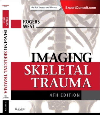
Imaging Skeletal Trauma
Seiten
2015
|
4th Revised edition
W B Saunders Co Ltd (Verlag)
978-1-4377-2779-1 (ISBN)
W B Saunders Co Ltd (Verlag)
978-1-4377-2779-1 (ISBN)
- Titel ist leider vergriffen;
keine Neuauflage - Artikel merken
Last published over a decade ago, this classic radiology text has been exhaustively updated by leading experts to provide the latest techniques and advances available in radiology today. Exceptional in scope and lavishly illustrated throughout, Imaging of Skeletal Trauma continues to offer a comprehensive view of diagnostic imaging in the evaluation of skeletal trauma, now in one consolidated single volume.
".. the change in focus from an authoritative reference to primer has been a good one.." Reviewed by RAD Magazine, June 2015
Master imaging techniques for the patient with multiple injuries, and understand the epidemiology and classification of various fractures, including chondral, osteochondral, stress, and pathologic.
Explore the effects of various traumatic childhood injuries on the growing skeleton.
Address the diagnostic pitfalls for a complete range of common, rare, and acute injuries.
Access up-to-date information on the role of helical CT and MR imaging in the evaluation of acute skeletal trauma.
View nearly 3,000 radiographs, CTs, and MR images, along with a wealth of line drawings that richly depict the principal features of all common fractures and dislocations.
Access the most important, need-to-know information regarding all aspects of imaging skeletal trauma with this consolidated single-volume edition.
Quickly reference critical material with an organization based on anatomical region.
Efficiently read and understand images while in an emergency setting with an expanded presentation of CT and MRI.
Take advantage of global expertise from brand-new contributing authors, including diagnostic radiologist Dr. O. Clark West.
View the fully searchable contents online at Expert Consult.
Chapter 1: Introduction
Chapter 2: The Shoulder
Chapter 3: The Elbow
Chapter 4: The Wrist
Chapter 5: The Hand
Chapter 6: The Cervical Spine
Chapter 7: The Thoracolumbar Spine
Chapter 8: The Pelvis
Chapter 9: The Hip
Chapter 10: The Knee
Chapter 11: The Ankle
Chapter 12: The Foot
| Erscheint lt. Verlag | 29.1.2015 |
|---|---|
| Zusatzinfo | Approx. 575 illustrations (75 in full color) |
| Verlagsort | London |
| Sprache | englisch |
| Maße | 222 x 281 mm |
| Themenwelt | Medizinische Fachgebiete ► Chirurgie ► Unfallchirurgie / Orthopädie |
| Medizin / Pharmazie ► Medizinische Fachgebiete ► Onkologie | |
| Medizin / Pharmazie ► Medizinische Fachgebiete ► Orthopädie | |
| Medizin / Pharmazie ► Medizinische Fachgebiete ► Radiologie / Bildgebende Verfahren | |
| ISBN-10 | 1-4377-2779-4 / 1437727794 |
| ISBN-13 | 978-1-4377-2779-1 / 9781437727791 |
| Zustand | Neuware |
| Haben Sie eine Frage zum Produkt? |
Mehr entdecken
aus dem Bereich
aus dem Bereich
Buch | Hardcover (2012)
Westermann Schulbuchverlag
34,95 €
Schulbuch Klassen 7/8 (G9)
Buch | Hardcover (2015)
Klett (Verlag)
30,50 €
Buch | Softcover (2004)
Cornelsen Verlag
25,25 €


