Point of Care Ultrasound
W B Saunders Co Ltd (Verlag)
978-1-4557-7569-9 (ISBN)
- Titel erscheint in neuer Auflage
- Artikel merken
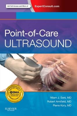
Access all the facts with focused chapters covering a diverse range of topics, as well as case-based examples that include ultrasound scans and videos.
Understand the pearls and pitfalls of point-of-care ultrasound through contributions from experts at more than 30 institutions.
View techniques more clearly than ever before. Illustrations and photos include transducer position, cross-sectional anatomy, ultrasound cross sections, and ultrasound images.
Expert Consult eBook version included with purchase. This enhanced eBook experience allows you to watch more than 200 ultrasound videos from real-life patients that demonstrate key findings; these videos are complemented by anatomical illustrations and text descriptions to maximize learning. You'll also be able to search all of the text, figures,�and references from the book on a variety of devices.
Section I: Fundamental Principles of Ultrasound
Chapter 1: Evolution of Point-of-Care Ultrasound
Chapter 2: Ultrasound Physics
Chapter 3: Transducers
Chapter 4: Orientation
Chapter 5: Basic Operation of an Ultrasound Machine
Chapter 6: Artifacts
Section II: Lung and Pleura
Chapter 7: Overview
Chapter 8: Lung and Pleural Ultrasound Technique
Chapter 9: Lung Ultrasound Interpretation
Chapter 10: Pleural Ultrasound Interpretation
Chapter 11: Lung and Pleural Procedures
Section III: Heart
Chapter 12: Overview
Chapter 13: Cardiac Ultrasound Technique
Chapter 14: Left Ventricular Function
Chapter 15: Right Ventricular Function
Chapter 16: Valves
Chapter 17: Pericardial Effusion
Chapter 18: Inferior Vena Cava
Section IV: Abdomen and Pelvis
Chapter 19: Gallbladder
Chapter 20: Kidneys
Chapter 21: Bladder
Chapter 22: Abdominal Aorta
Chapter 23: Abdominal Fluid
Chapter 24: First Trimester Pregnancy and Normal Female Reproductive System
Chapter 25: Testicular Ultrasound
Section V: Vascular System
Chapter 26: Lower Extremity Deep Venous Thrombosis
Chapter 27: Upper Extremity Deep Venous Thrombosis
Chapter 28: Central Venous Access
Chapter 29: Peripheral Venous Access
Chapter 30: Arterial Access
Section VI: Head and Neck
Chapter 31: Ocular Ultrasound
Chapter 32: Thyroid Gland
Chapter 33: Lymph Nodes
Section VII: Nervous System
Chapter 34: Peripheral Nerve Blocks
Chapter 35: Lumbar Puncture
Section VIII: Soft Tissues and Joints
Chapter 36: Skin and Soft Tissues
Chapter 37: Joints
Section IX: Clinical Scenarios and Protocols
Chapter 38: Dyspnea and Acute Respiratory Failure
Chapter 39: Abdominal Pain
Chapter 40: Hypotension and Shock
Chapter 41: Trauma
Chapter 42: Cardiac Arrest
Section X: Ultrasound Program Management
Chapter 43: Competence, Credentialing, and Certification
Chapter 44: Equipment, Image Archiving, and Billing
Index
| Zusatzinfo | Approx. 100 illustrations (100 in full color) |
|---|---|
| Verlagsort | London |
| Sprache | englisch |
| Maße | 152 x 229 mm |
| Themenwelt | Medizinische Fachgebiete ► Radiologie / Bildgebende Verfahren ► Sonographie / Echokardiographie |
| ISBN-10 | 1-4557-7569-X / 145577569X |
| ISBN-13 | 978-1-4557-7569-9 / 9781455775699 |
| Zustand | Neuware |
| Informationen gemäß Produktsicherheitsverordnung (GPSR) | |
| Haben Sie eine Frage zum Produkt? |
aus dem Bereich


