Atlas of Orthopaedic Radiology
Lippincott Williams and Wilkins (Verlag)
978-1-56375-023-6 (ISBN)
- Titel erscheint in neuer Auflage
- Artikel merken
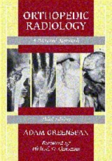
Directed at the needs of orthopaedists and residents in orthopaedics, as well as radiologists and radiologists in training, this book aims to facilitate the complex process of diagnostic investigation. Clearly, systematically and logically, it strives to provide basic understanding of contemporary imaging methods, such as radiography, scanography, arthrography, scintigraphy, stress views, digital radiography, computed tomography, MRI, myelography and sonography. It analyzes and demonstrates the comparative diagnostic utility of different imaging studies in different settings, including trauma osteoarthritis, rheumatoid arthritis, tumours and tumour-like lesions, infections, metabolic and endocrine disorders, and congenital and developmental anomalies. Further, the work offers guidance in selecting the most effective radiological techniques - aimed at minimizing cost and radiation exposure - in specific settings. The second edition features two new chapters: one on the application of different radiological imaging techniques in the evaluation of musculoskeletal disorders; the other concerns bone formation and growth.
There is also an updated and revised trauma section with numerous new illustrations, including innovative circular diagrams on imaging techniques for evaluating injury in various regions of the axial and appendicular skeleton. The second edition also covers the arthridites, with recent data on hyperuricaemia, rheumatoid factors, antinuclear antibodies, and synovial fluid analysis. These are accompanied by new sections on polymyositis, dermatomyositis, mixed connective tissue disease and systemic lupus erythematosus, among others. Finally, this edition contains key histopathological findings, and their clinical implications, in musculoskeletal tumours.
Part 1 Introduction to orthopaedic radiology: the role of the orthopaedic radiologist; imaging techniques in orthopaedics; growth and development of bones. Part 2 Trauma: radiologic evaluation of trauma; upper limb - shoulder girdle, elbow, distal forearm, wrist, hand; lower limb - pelvic girdle, proximal femur, knee, ankle, foot; spine. Part 3 Arthritides: radiologic evaluation of arthritides; degenerative joint diseases; inflammatory arthritides; miscellaneous arthritides. Part 4 Tumours and tumour-like lesions: radiologic evaluation of tumours and tumour-like lesions; benign tumours and tumour-like lesions - osteoblastic and chondroblastic lesions, fibrous, fibro-osseous, fibrohistiocytic lesions and other benign conditions; malignant bone tumours. Part 5 Infections: radiologic evaluation of musculoskeletal infections; osteomyelitis, infectious arthritis and soft tissue infections. Part 6 Metabolic and endocrine disorders: radiologic evaluation of metabolic and endocrine disorders; osteoporosis, rickets and osteomalacia; hyperparathyroidism; Paget's disease; miscellaneous metabolic and endocrine disorders. Part 7 Congenital and developmental anomalies: radiologic evaluation of skeletal anomalies; anomalies of the upper and lower limbs; scoliosis.
| Erscheint lt. Verlag | 1.9.1992 |
|---|---|
| Zusatzinfo | 1200 illustrations |
| Verlagsort | Philadelphia |
| Sprache | englisch |
| Maße | 253 x 307 mm |
| Gewicht | 3580 g |
| Themenwelt | Medizinische Fachgebiete ► Chirurgie ► Unfallchirurgie / Orthopädie |
| Medizinische Fachgebiete ► Radiologie / Bildgebende Verfahren ► Kernspintomographie (MRT) | |
| Medizinische Fachgebiete ► Radiologie / Bildgebende Verfahren ► Radiologie | |
| ISBN-10 | 1-56375-023-6 / 1563750236 |
| ISBN-13 | 978-1-56375-023-6 / 9781563750236 |
| Zustand | Neuware |
| Haben Sie eine Frage zum Produkt? |
aus dem Bereich


