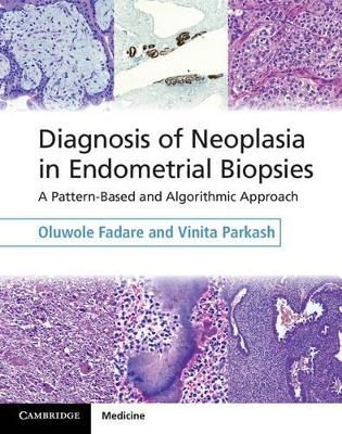
Diagnosis of Neoplasia in Endometrial Biopsies Book and Online Bundle
Cambridge University Press
978-1-107-04043-4 (ISBN)
With its unique algorithmic and pattern-based approach, Diagnosis of Neoplasia in Endometrial Biopsies is an essential practical guide to interpreting endometrial biopsy samples. All potential entities are classified based on the dominant histologic pattern, with each resulting sub-group progressively sub-classified to reach a diagnosis. Decision tree flowcharts facilitate rapid narrowing of the differential diagnosis. Recent advancements are discussed and explained, and strengths and limitations of diagnostic tests are identified in the context of their application to the biopsy sample. Lavishly illustrated throughout, this book serves the practising pathologist as a scope-side assistant for quick reference, up-to-date guidance, and recommendations for ancillary testing. For the resident, this book facilitates quick and comprehensive mastery of the interpretation and diagnosis of endometrial biopsies. The book is packaged with a password, giving the user online access to all the text and images.
Dr Oluwole Fadare, MD is an Associate Professor of Pathology, Microbiology and Immunology and of Obstetrics and Gynecology at Vanderbilt University School of Medicine, and Team Leader for Gynecologic Pathology, Associate Director of Surgical Pathology, and Medical Director of the Immunohistochemistry Laboratory at Vanderbilt University Medical Center, Nashville, TN, USA. Dr Vinita Parkash, MD is an Associate Professor of Pathology at Yale University School of Medicine, New Haven, and the Director of Surgical Pathology at Bridgeport Hospital, Bridgeport, CT, USA.
Preface; 1. General principles in the evaluation of endometrial samples; 2. Endometrial samples with roughly equal ratio of glands to stroma; 3. Lesions with epithelium to stroma ratio in excess of 1:1; 4. Purely epithelial proliferations (no significant stromal component); 5. Spindle cell and myxoid lesions; 6. Round cell lesions; 7. Epithelioid cell lesions; 8. Trophoblastic and gestational lesions; Index.
| Erscheint lt. Verlag | 4.9.2014 |
|---|---|
| Zusatzinfo | 15 Tables, color; 374 Halftones, color; 19 Line drawings, color |
| Verlagsort | Cambridge |
| Sprache | englisch |
| Maße | 222 x 282 mm |
| Gewicht | 900 g |
| Themenwelt | Medizin / Pharmazie ► Medizinische Fachgebiete ► Onkologie |
| Studium ► 2. Studienabschnitt (Klinik) ► Pathologie | |
| ISBN-10 | 1-107-04043-4 / 1107040434 |
| ISBN-13 | 978-1-107-04043-4 / 9781107040434 |
| Zustand | Neuware |
| Haben Sie eine Frage zum Produkt? |
aus dem Bereich

