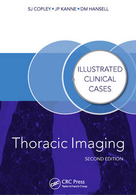
Thoracic Imaging
Illustrated Clinical Cases, Second Edition
Seiten
2014
|
2nd edition
Crc Press Inc (Verlag)
978-1-4822-3115-1 (ISBN)
Crc Press Inc (Verlag)
978-1-4822-3115-1 (ISBN)
- Titel z.Zt. nicht lieferbar
- Versandkostenfrei innerhalb Deutschlands
- Auch auf Rechnung
- Verfügbarkeit in der Filiale vor Ort prüfen
- Artikel merken
The chest radiograph is a ubiquitous, first-line investigation and accurate interpretation is often difficult. Radiographic findings may lead to the use of more sophisticated imaging techniques, such as multidetector computed tomography (MDCT) and positive emission tomography.
Containing 100 challenging clinical cases and illustrated with superb, high quality images, Thoracic Imaging, Second Edition explores a wide range of lung conditions. Coverage ranges from basic radiographic cases such as tuberculosis, pulmonary and mediastinal masses to the more challenging diseases, including cystic fibrosis, asbestosis, sarcoidosis and interstitial lung disease.
It remains an invaluable text for radiologists and imaging professionals in practice and in training, from hospital-based doctors preparing for higher examinations to established physicians in their continuing professional development.
Sue Copley, David Hansell, Jeffrey Kanne
Preface. List of Abbreviations. Glossary. Clinical Cases. Index.
| Erscheint lt. Verlag | 4.6.2014 |
|---|---|
| Reihe/Serie | Illustrated Clinical Cases |
| Zusatzinfo | 238 Illustrations, color |
| Verlagsort | Bosa Roca |
| Sprache | englisch |
| Maße | 174 x 246 mm |
| Gewicht | 424 g |
| Themenwelt | Medizinische Fachgebiete ► Innere Medizin ► Kardiologie / Angiologie |
| Medizinische Fachgebiete ► Innere Medizin ► Pneumologie | |
| Medizinische Fachgebiete ► Radiologie / Bildgebende Verfahren ► Radiologie | |
| ISBN-10 | 1-4822-3115-8 / 1482231158 |
| ISBN-13 | 978-1-4822-3115-1 / 9781482231151 |
| Zustand | Neuware |
| Haben Sie eine Frage zum Produkt? |
Mehr entdecken
aus dem Bereich
aus dem Bereich
in Fällen, Fragen und Antworten
Buch | Softcover (2024)
Urban & Fischer in Elsevier (Verlag)
89,00 €
Diagnostik und interventionelle Therapie | 2 Bände
Buch (2024)
Deutscher Ärzteverlag
349,99 €


