Atlas of Tissue Doppler Echocardiography — TDE
Steinkopff (Verlag)
978-3-642-47069-1 (ISBN)
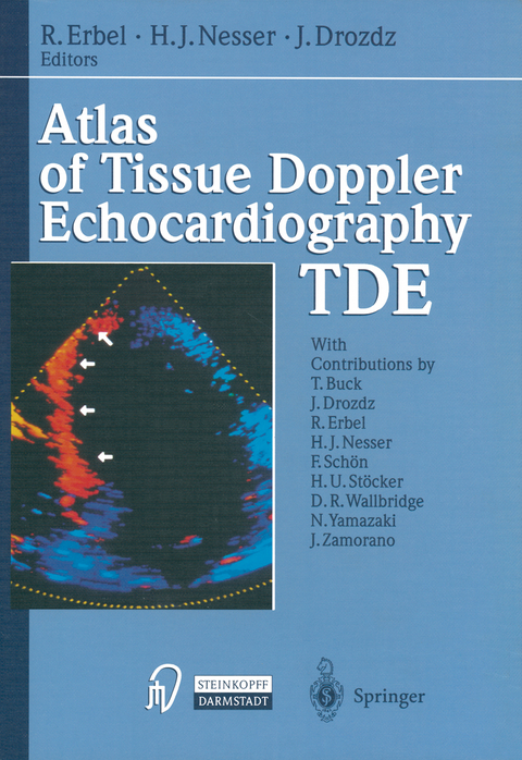
This is the first book to present an overview of the exciting new cardiac imaging technique of tissue Doppler echocardiography (TDE). In order to understand the background of this technique, it is necessary to compare the physical properties of blood, which reflects ultrasound poorly but moves with high velocity (up to 150 cm/s) with those of the myocar dium, which reflects ultrasound strongly but moves with low velocity (less than 10 cm/s). In tissue Doppler imaging, existing Doppler technology has been modified to bypass the high-pass filter and enhance calculation of low velocities, thus enabling selective visualization of the myocardium rather than of the blood. Because the color Doppler tissue images are super imposed on the conventional two-dimensional ultrasound images, this technique is known as TDE. Following a brief introduction, the history of ultrasound and Doppler imaging is presented. It is now about 150 years since the death of Christian Doppler, who described the "Doppler" effect, and more than 100 years since Pierre Curie discovered the piezoelectric effects of crystals. TDE was developed by Nobuo Yamazaki and Yoshitaka Mine at the Medi cal Engineering Laboratory, Toshiba Corporation, Tochigi, Japan. En gineers involved in the development of the technique have provided important technical information, which the reader will find an invaluable background to potential applications ofTDE.
List of Contents.- 1 Introduction.- 2 Milestones in Cardiovascular Ultrasound.- 3 Principle of Doppler Tissue Velocity Measurements.- 4 Image Processing.- 5 Normal Pattern of Myocardial Velocity.- 6 Movement of the Total Heart.- 7 Assessment of Right Ventricular Wall Thickness by Tissue Doppler Echocardiography.- 8 Ischemic Heart Disease.- 9 TDE and Stress Echocardiography.- 10 Hypertrophic Cardiomyopathy.- 11 Cardiac Amyloidosis.- 12 Aortic Wall Velocity.- 13 Hemodynamics by TDE.- 14 Miscellaneous.- Perspectives.
Pressestimmen: "Erbel und Mitarbeiter haben einen Atlas vorgelegt, der einzigartig ist und neue Erkenntnisse hinsichtlich Textur, Morphologie und Funktion des Herzmuskels aufzeigt. ... Diese neue Methode ermöglicht somit neue diagnostische Ansätze, aber auch neue Möglichkeiten zur Therapiekontrolle.
Wenngleich das Buch für den Experten gedacht ist, wird auch der Arzt, der nicht Kardiologe ist, diesen Atlas mit Muße und Gewinn studieren können."
(Deutsches Ärzteblatt)
| Erscheint lt. Verlag | 23.2.2012 |
|---|---|
| Zusatzinfo | IX, 169 p. 101 illus. in color. |
| Verlagsort | Heidelberg |
| Sprache | englisch |
| Maße | 210 x 279 mm |
| Gewicht | 453 g |
| Themenwelt | Medizinische Fachgebiete ► Innere Medizin ► Kardiologie / Angiologie |
| Medizin / Pharmazie ► Studium | |
| Schlagworte | Assessment • Cardiovascular • clinical application • Echocardiography • heart • Heart disease • hemodynamics • tissue • Ultrasound |
| ISBN-10 | 3-642-47069-6 / 3642470696 |
| ISBN-13 | 978-3-642-47069-1 / 9783642470691 |
| Zustand | Neuware |
| Haben Sie eine Frage zum Produkt? |
aus dem Bereich


