


Cone-beam x-ray phase contrast tomography of biological samples
Optimization of contrast, resolution and field of view
Seiten
2013
Universitätsverlag Göttingen
978-3-86395-134-4 (ISBN)
Universitätsverlag Göttingen
978-3-86395-134-4 (ISBN)
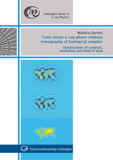
Three-dimensional information of entire objects can be obtained by the remarkable technique of computed tomography (CT). In combination with phase sensitive X-ray imaging high contrast for soft tissue structures can be achieved as opposed to CT based on classical radiography. In this work biological samples ranging from micrometer sized single cells over multi-cellular nerve tissue to entire millimeter sized organs are investigated by use of cone-beam propagationbased X-ray phase contrast. Optimization with respect to contrast, resolution and field of view is achieved by addressing instrumentation, sample preparation and phase reconstruction techniques. By using laboratory sources functional soft tissue within the bony capsule of mouse cochleae is visualized in 3D with unprecedented image quality. At synchrotron storage rings the technique reveals more than 1000 axons running in parallel within a mouse nerve and enables doseefficient three-dimensional cellular imaging as well as two-dimensional imaging at high resolutions below 50 nm.
| Reihe/Serie | Göttingen Series in X-ray Physics ; 13 |
|---|---|
| Sprache | englisch |
| Maße | 170 x 240 mm |
| Einbandart | Paperback |
| Themenwelt | Medizinische Fachgebiete ► Radiologie / Bildgebende Verfahren ► Nuklearmedizin |
| Naturwissenschaften ► Physik / Astronomie | |
| Schlagworte | Computed tomography • radiography • X-ray physics |
| ISBN-10 | 3-86395-134-4 / 3863951344 |
| ISBN-13 | 978-3-86395-134-4 / 9783863951344 |
| Zustand | Neuware |
| Haben Sie eine Frage zum Produkt? |
Mehr entdecken
aus dem Bereich

aus dem Bereich
Buch | Softcover (2022)
Lehmanns Media (Verlag)
39,95 €
Buch | Softcover (2022)
Facultas (Verlag)
31,10 €