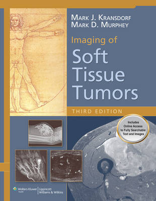
Imaging of Soft Tissue Tumors
Seiten
2013
|
3rd edition
Lippincott Williams and Wilkins (Verlag)
978-1-4511-1641-0 (ISBN)
Lippincott Williams and Wilkins (Verlag)
978-1-4511-1641-0 (ISBN)
- Titel ist leider vergriffen;
keine Neuauflage - Artikel merken
Plays an essential role in the diagnosis of soft tissue tumors as well as in surgical planning. Based on cases seen at the Armed Forces Institute of Pathology and the Mayo Clinic, this book offers detailed visually supported information on the radiologic evaluation of soft tissue tumors and tumor-like lesions.
Imaging of Soft Tissue Tumors, Third Edition
Imaging technology plays an essential role in the diagnosis of soft tissue tumors as well as in surgical planning. Not only can imaging studies such as CT and MRI determine the relationship between a tumor and adjacent vessels and nerves, but, because some soft tissue tumors possess specific radiologic presentations, imaging can help pinpoint the tumor type.
Based on cases seen at the Armed Forces Institute of Pathology and the Mayo Clinic, this comprehensive reference offers detailed visually supported information on the radiologic evaluation of soft tissue tumors and tumor-like lesions. Inside, readers will explore the full spectrum of soft tissue pathologies, with over 1400 images that highlight both common and atypical presentations. The book’s expert authors offer valuable advice on selecting the most appropriate imaging modality for each tumor type.
Features:
Broad scope presents the results of a retrospective analysis of over 31,000 cases of soft tissue tumors and tumor-like masses.
Comprehensive coverage addresses lipomatous, vascular and lymphatic, fibrous and fibrohistiocytic, muscle, neurogenic, synovial, extraskeletal osseous and cartilaginous tumors.
Detailed information on all imaging modalities —including CT, MRI, radiography, angiography, scintigraphy, ultrasound, and positron emission tomography—are covered, with facts on the strengths and limitations of each to help readers select the most appropriate method.
High-quality images aid in the visual diagnosis of typical and atypical tumor presentations.
World Health Organization nomenclature (in accordance with the 2013 updated classification) is included throughout the text, emphasizing applicable changes in each chapter.
A new chapter examines the imaging of superficial masses.
| Erscheint lt. Verlag | 25.10.2013 |
|---|---|
| Zusatzinfo | 1510 |
| Verlagsort | Philadelphia |
| Sprache | englisch |
| Maße | 213 x 276 mm |
| Gewicht | 2132 g |
| Themenwelt | Medizin / Pharmazie ► Medizinische Fachgebiete ► Onkologie |
| Medizin / Pharmazie ► Medizinische Fachgebiete ► Radiologie / Bildgebende Verfahren | |
| ISBN-10 | 1-4511-1641-1 / 1451116411 |
| ISBN-13 | 978-1-4511-1641-0 / 9781451116410 |
| Zustand | Neuware |
| Haben Sie eine Frage zum Produkt? |
Mehr entdecken
aus dem Bereich
aus dem Bereich
Buch | Hardcover (2012)
Westermann Schulbuchverlag
34,95 €
Schulbuch Klassen 7/8 (G9)
Buch | Hardcover (2015)
Klett (Verlag)
30,50 €
Buch | Softcover (2004)
Cornelsen Verlag
25,25 €


