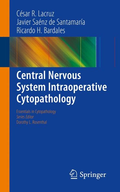
Central Nervous System Intraoperative Cytopathology
Springer-Verlag New York Inc.
978-1-4614-8428-8 (ISBN)
- Titel erscheint in neuer Auflage
- Artikel merken
The Essentials in Cytopathology book series fulfills the need for an easy-to-use and authoritative synopsis of site specific topics in cytopathology. These guide books fit into the lab coat pocket and are ideal for portability and quick reference. Each volume is heavily illustrated with a full color art program, while the text follows a user-friendly outline format.
Central Nervous System Intraoperative Cytopathology covers the full spectrum of benign and malignant conditions of the CNS with emphasis on common disorders. The volume is heavily illustrated and contains useful algorithms that guide the reader through the differential diagnosis of common and uncommon entities encountered in the field of intraoperative neuro-cytopathology. Central Nervous System Intraoperative Cytopathology is a valuable quick reference for pathologists, cytopathologists, and fellows and trainees dealing with this exigent field.
César R. Lacruz, MD, PhD, FIAC Consultant Pathologist, Assistant Professor Pathology University General Hospital “Gregorio Marañón”, Department of Pathology, Madrid, Spain Javier Saénz de Santamaría, MD, PhD, FIAC Director of Anatomic Pathology, Professor of Pathology University Hospital, Extremadura Medical School, Badajoz, Spain Ricardo H. Bardales, MD, MIAC Staff Pathologist Outpatient Pathology Associates, Sacramento, CA, USA
Chapter 1 Introduction to CNS Intraoperative Cytopathology
Historical Background
Accuracy of CNS Intraoperative Cytopathology
Histologic Types of CNS Neoplasms
Chapter 2 Clinical, Radiologic, and Technical Considerations
Clinical Considerations
Radiologic Considerations
Technical Considerations
Chapter 3 Algorithmic Approach to CNS Intraoperative Cytopathology
Sample Triage
Smear Evaluation
General Category Interpretation
Chapter 4 Normal Brain and Gliosis
White Matter Pattern
Gray Matter Pattern
Cerebellar Cortex Pattern
Choroid Plexus Pattern
Leptomeningeal Pattern
Reactive gliosis
Contaminants
Chapter 5 Astrocytic Tumors
Diffusely Infiltrating Astrocytomas
Diffuse Astrocytoma
Anaplastic Astrocytoma
Glioblastoma
Pilocytic astrocytoma
Special Astrocytic Tumors
Subependymal Giant Cell Astrocytoma
Pleomorphic Xanthoastrocytoma
Gliomatosis Cerebri
Chapter 6 Oligodendroglial Tumors
Oligodendroglioma
Anaplastic Oligodendroglioma
Chapter 7 Ependymal Tumors
Ependymoma
Anaplastic Ependymoma
Subependymoma
Myxopapillary Ependymoma
Chapter 8 Choroid Plexus Tumors
Choroid Plexus Papilloma
Choroid Plexus Carcinoma
Chapter 9 Neuronal and Glioneural Tumors
Desmoplastic Infantile Ganglioglioma/Astrocytoma
Dysembryoplastic Neuroepithelial Tumor
Gangliocytoma and Ganglioglioma
Central Neurocytoma
Spinal Paraganglioma
Chapter 10 Embryonal Tumors
Medulloblastoma
Primitive Neuroectodermal Tumors
Atypical Teratoid-Rhabdoid Tumor
Chapter 11 Meningeal Tumors
Meningioma
Hemangioblastoma
Hemangiopericytoma
Chapter 12 CNS Germ Cell Tumors
Germinoma
Teratomas
Malignant Non-germinomatous Germ Cell Tumors
Chapter 13 Tumors of the Hematopoietic System
Primary CNS Lymphoma
Plasmacytoma
Granulocytic Sarcoma
Histiocytic Lesions
Chapter 14 Tumors of the Cranial and Spinal Nerves
Schwannoma
Neurofibroma
Chapter 15 Tumors of the Pineal Region
Pineocytoma
Pineoblastoma
Pineal Glial Cyst
Chapter 16 Tumors of the Sellar Region
Pituitary Adenoma
Craniopharyngioma
Other Lesions of the Sellar Region
Chapter 17 Metastatic Tumors
Chapter 18 Benign Cystic Lesions
Squamous Epithelium-Lined Cysts
Columnar Epithelium-Lined Cysts
Non Epithelial-Lined Cysts
Chapter 19 Non-Neoplastic Disorders
Acute Inflammatory Cell-Rich Lesions
Epithelioid-Cell and Lymphoid-Cell Rich Lesions
Macrophage-Rich Lesions
Inflammatory Lesions in AIDS
Chapter 20 Extradural Mass Lesions Compressing the Spinal Cord
Neoplastic Lesions
Non Neoplastic Lesions
| Reihe/Serie | Essentials in Cytopathology ; 13 |
|---|---|
| Zusatzinfo | 46 Tables, black and white; 129 Illustrations, color; XIV, 276 p. 129 illus. in color. |
| Verlagsort | New York, NY |
| Sprache | englisch |
| Maße | 127 x 204 mm |
| Gewicht | 3546 g |
| Themenwelt | Medizin / Pharmazie ► Medizinische Fachgebiete ► Laboratoriumsmedizin |
| Studium ► 2. Studienabschnitt (Klinik) ► Pathologie | |
| Schlagworte | Choroid plexus • Glioneural tumors • Hemangioblastoma • Hematopoietic tissue • Meningioma • Neuroglia |
| ISBN-10 | 1-4614-8428-6 / 1461484286 |
| ISBN-13 | 978-1-4614-8428-8 / 9781461484288 |
| Zustand | Neuware |
| Informationen gemäß Produktsicherheitsverordnung (GPSR) | |
| Haben Sie eine Frage zum Produkt? |
aus dem Bereich
