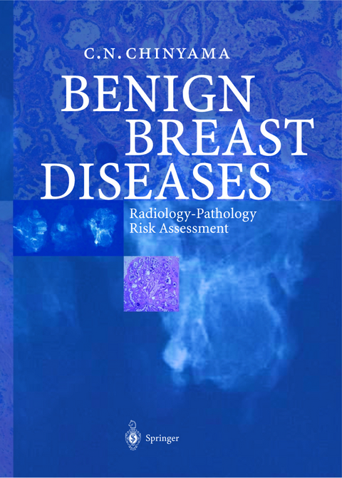
Benign Breast Diseases
Springer Berlin (Verlag)
978-3-642-62146-8 (ISBN)
The majority of textbooks on breast disease understandably concentrate on cancer and related prognostic factors, with minimal space devoted to benign conditions. With widespread use of screening mammography and improvements in imaging equipment, smallerand smaller cancers are being detected. This has also resulted in detection of indeterminate micro-calcification and soft tissue densities, which in variablyleads to diagnostic fine needle aspirationcytologyor needle core biopsy. Al though most of the mammographically indeterminate lesions are benign, biopsies also detect potentially malignant lesions, which include atypical hyperplasias, le sions of undetermined malignant potential such as columnar cell change and other epithelial proliferations. These lesions require proper radiological and pathological assessment at multidisciplinary meetings for appropriate patient management. The objective ofthis bookisto provide an overview ofradiological and pathologi cal features of benign lesions with an emphasis on screen-detected lesions, with il lustrated examples. Although the radiologicaland pathological correlations concen trate on the screen-detectedbenignlesions, it wasnot possible to limit the discussion of the radiologyand pathologyto justscreen-detectedlesions. This isbecause mam mographic screeningbecameroutine in developed countries only in the past fewde cades, and there are insufficient follow-up data in the literature on patients with screen-detectedbenign disease. Secondly,although breast disease isarbitrarilyclas sified into symptomatic and screen-detected, all women who attend specialised breast units invariablyundergo some form ofimaging, mostly mammographyor ul trasound. Emphasis on screen-detectedlesions also highlights the heterogeneous ar ray of benign diseases in this age group, which in the future may create significant breast disease workloads as longevity becomes the norm in the developed world.
Dr. Chinyama qualified with Honours Degree in Medicine in Harare, Zimbabwe, Trained in Breast Pathology at St. Bartholomew's Hospital, London and Bristol South West Breast Screening Unit in Bristol,UK. Worked as Senior Lecturer/Honorary Consultant in Histopathology at Guy's and St.Thomas' Hospital, London. Currently working as a Consultant Pathologist, Princess Elizabeth Hospital, Guernsey, Channel Islands.
1 Radiology of Benign Breast Disease.- 2 Surgery of Benign Breast Disease.- 3 Pathology of Benign Breast Disease.- 4 Fibre-epithelial Lesions.- 5 Infiltrative Pseudo-malignant Lesions.- 6 Hyperplastic Epithelial Lesions.- 7 Cystic Lesions.- 8 Mucocele-like Lesions.- 9 Columnar Cell Lesions.- 10 Calcification in Benign Lesions.- 11 Non-epithelial Lesions.- 12 Risk Assessment in Benign Breast Disease.
From the reviews:
"The advent of breast screening ... now means that the breast team is regularly presented with a variety of benign pathological diagnoses and an understanding of the clinical relevance of these is essential. This text aims to contribute towards this understanding. ... Pathological slides are beautifully reproduced in full colour ... . a comprehensive text on the pathology and risk assessment of benign breast disease which is well illustrated with pathological slides. ... it would represent useful supplementary reading for non-pathologists ... ." (Dr. J Litherland, RAD Magazine, July, 2005)
"Catherine Chinyama has made a creditable attempt to bring order to the often confusing field of benign breast disease. ... The main focus of the book is on radiologic and pathological findings in benign breast disease ... . The book is remarkably well illustrated ... . The figures are informative ... . The range of benign breast disease covered in the book is broad ... . thisvery useful book will interest any clinician, radiologist, or pathologist who deals with benign breast disease ... ." (Pamela J. Goodwin, The New England Journal of Medicine, September, 2004)
| Erscheint lt. Verlag | 5.11.2012 |
|---|---|
| Zusatzinfo | XIV, 150 p. |
| Verlagsort | Berlin |
| Sprache | englisch |
| Maße | 193 x 270 mm |
| Gewicht | 378 g |
| Themenwelt | Medizinische Fachgebiete ► Radiologie / Bildgebende Verfahren ► Radiologie |
| Schlagworte | Benign • Breast Disease • Pathology • Radiology • Risk • Surgery |
| ISBN-10 | 3-642-62146-5 / 3642621465 |
| ISBN-13 | 978-3-642-62146-8 / 9783642621468 |
| Zustand | Neuware |
| Informationen gemäß Produktsicherheitsverordnung (GPSR) | |
| Haben Sie eine Frage zum Produkt? |
aus dem Bereich


