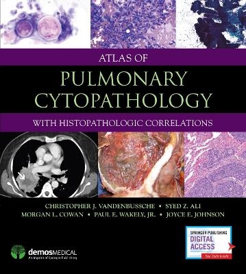
Atlas of Pulmonary Cytopathology
Seiten
2017
Demos Medical Publishing (Verlag)
978-1-936287-16-1 (ISBN)
Demos Medical Publishing (Verlag)
978-1-936287-16-1 (ISBN)
- Titel z.Zt. nicht lieferbar
- Versandkostenfrei innerhalb Deutschlands
- Auch auf Rechnung
- Verfügbarkeit in der Filiale vor Ort prüfen
- Artikel merken
Offers concrete diagnostic guidance for anatomic pathologists to accurately identify pulmonary disease using exfoliative and aspiration techniques. It not only illustrates the cytomorphology of lung specimens, but also presents and contrasts common problem areas that can lead to erroneous interpretation.
Atlas of Pulmonary Cytopathology: With Histopathologic Correlations offers concrete diagnostic guidance for anatomic pathologists to accurately identify pulmonary disease using exfoliative and aspiration techniques. It not only illustrates the cytomorphology of lung specimens, but also presents and contrasts common problem areas that can lead to erroneous interpretation. Clearly and concisely written by leaders in the field, this volume is a practical desk reference for all facets of the diagnostically challenging area of pulmonary cytopathology.
The Atlas features nearly 500 carefully selected high-resolution color images detailing important aspects of the full range of lung diseases and conditions including infections, non-neoplasic disorders, benign neoplasms, and malignant tumors such as adenocarcinoma, squamous cell carcinoma, neuroendocrine tumors, epithelial tumors and malignant mesothelioma, mesenchymal and lymphohistiocytic tumors, as well as metastatic tumors. Additionally, the book's images of the histopathology and gross characteristics of lesions provide morphologic correlations that will be relevant to cytopathologists and surgical pathologists alike. To provide a broader, more enriching perspective, the Atlas features a special chapter on the bronchoscopic characteristics of lung lesions to provide a differential diagnosis through the eyes of an experienced pulmonologist. It also reviews the updated classification guidelines of lung tumors from the World Health Organization (WHO), providing a multidisciplinary approach to enhance the reader's understanding of how cytopathology, histopathology, and bronchoscopic information together create a powerful tool for the prevention and early detection of neoplastic, non-neoplastic, and infectious disease of the lower respiratory tract.
Key Features:
Provides practical, expert diagnostic guidance for the full range of pulmonary cytopathology
Illuminates common diagnostic pitfalls and interpretive errors made in lower respiratory tract specimens
Presents nearly 500 high-resolution color images
Includes bronchoscopic-pathologic correlations from an expert pulmonologist
| Erscheint lt. Verlag | 30.11.2017 |
|---|---|
| Zusatzinfo | 500 Illustrations |
| Verlagsort | New York, NY |
| Sprache | englisch |
| Maße | 229 x 254 mm |
| Gewicht | 454 g |
| Themenwelt | Medizinische Fachgebiete ► Chirurgie ► Herz- / Thorax- / Gefäßchirurgie |
| Medizinische Fachgebiete ► Innere Medizin ► Pneumologie | |
| Studium ► 2. Studienabschnitt (Klinik) ► Pathologie | |
| ISBN-10 | 1-936287-16-1 / 1936287161 |
| ISBN-13 | 978-1-936287-16-1 / 9781936287161 |
| Zustand | Neuware |
| Informationen gemäß Produktsicherheitsverordnung (GPSR) | |
| Haben Sie eine Frage zum Produkt? |
Mehr entdecken
aus dem Bereich
aus dem Bereich
Diagnostische und interventionelle Kathetertechniken
Buch (2022)
Thieme (Verlag)
220,00 €
Buch | Hardcover (2022)
Urban & Fischer in Elsevier (Verlag)
270,00 €


