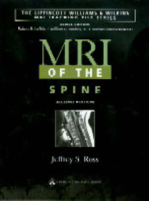
MRI of the Spine
Lippincott Williams and Wilkins (Verlag)
978-0-7817-2528-6 (ISBN)
- Titel ist leider vergriffen;
keine Neuauflage - Artikel merken
The thoroughly revised and updated Second Edition of this text is part of the popular Lippincott Williams & Wilkins MRI Teaching File Series . The book presents 100 actual case studies that cover a wide range of spinal disorders and demonstrate the use of current MRI techniques to aid in diagnosis.Each case study is illustrated with high-resolution MR images and presented in an easy-to-follow format on a two-page spread. On the left-hand page are the images and the clinical history. On the right-hand page are concise descriptions of the radiographic findings, the diagnosis, and the pathology. This format is ideal for teaching readers how to interpret MR images or for everyday reference at the view box.
Chiari II with Cervical Syrinx 2
Sacral Meningeal Cyst, Type I 4
Dermal Sinus with Tethered Cord 6
Diastematomyelia (Single Dura Investment) 8
Diastematomyelia with Spur 10
Epidermoid Tumor 12
Unilateral Hypoplastic Facet with Compensatory Contralateral Hypertrophy 14
Myelomeningocele 16
Caudal Regression 18
Arachnoiditis 20
Lumbar Canal Stenosis 22
Cervical Disk Herniation 24
Cervical Spondylosis with Focal Myelomalacia 26
Degenerative End-Plate Change Mimics Disk Space Infection 30
Lumbar Herniation with Enhancing Intradural Nerve 32
Type II Limbus Fracture with Herniation 36
Lumbar Canal Stenosis with Intradural Enhancement 38
Free Disk Fragment 40
Disk Extrusion 44
Free Disk Fragment 46
Ossification of the Posterior Longitudinal Ligament (OPLL) 48
Ossification of the Ligamentum Flavum with Cord Compression and Focal Myelomalacia 50
Postoperative Lumbar Hematoma 52
Lumbar Pseudoarthrosis 54
Pseudomeningocele 56
Disk Herniation and Epidural Fibrosis 58
Epidural Fibrosis 62
Scheuermann's Disease 64
Synovial Cyst 66
Ossified Thoracic Disk Herniation 68
Lateral Lumbar Disk Herniation 70
Type I>Cervical Cord Contusion in Cervical Spondylosis 74
Aspergillus Meningitis 78
Epidural Phlegmon 80
Demyelinating Disease (Multiple Sclerosis) 82
Postoperative Disk Space Infection 84
Disk Space Infection 86
Epidural Abscess with Meningitis 88
Multiple Sclerosis 90
Hereditary Motor and Sensory Neuropathy (Charcot-Marie-Tooth Disease) 92
Postinfectious Myelitis Involving Conus 94
Rheumatoid Arthritis 96
Sarcoidosis 98
Septic Facet Joint 100
Tuberculous Osteomyelitis/Spondylitis 102
Guillain-Barré Syndrome 104
Spinal Cysticercosis 106
Intramedullary Abscess 108
Multiple Sclerosis 110
Sarcoidosis 114
Tuberculous Spondylitis 116
Demyelinating Disease 118
Early Disk Space Infection Masked by Degenerative End-Plate and Disk Disease 122
Paget's Disease 124
Subacute Combined Degeneration 126
Idiopathic Spinal Cord Herniation 128
Glioma 130
Chordoma 132
Ependymoma 134
Ependymoma 136
Cord and Leptomeningeal Metastases 138
Melanocytoma 140
Meningioma 142
Myelomatous Meningitis 144
Neurofibromatosis, Type I 146
Neurofibromatosis, Type II 150
Osteoid Osteoma 152
Paraganglioma 154
Schwannoma 156
Schwannoma 158
Hemangioblastomas in Von Hippel-Lindau Syndrome 160
Multiple Myeloma 162
Meningioma 164
Cord Metastasis from Renal Cell Carcinoma 166
Leptomeningeal Metastasis 168
Superior Sulcus Tumor 170
Thoracic Ependymoma 172
Leptomeningeal Metastasis 176
Multiple Myeloma Following Vertebroplasty 178
Cavernous Angioma 180
Cord Hemorrhage Secondary to Vascular Malformation 182
Cord Infarct 184
Cord Infarct 186
Dural Fistula 188
Dural Fistula 190
Vertebral Body Hemangiomas 192
Superficial Siderosis 194
Subdural Hemorrhage 196
Epidural and Subdural Hemorrhage 198
Acute Epidural Hemorrhage 200
Os Odontoideum 202
L4 Spondylolysis 204
Right L3 Pars Interarticularis Fracture with Sclerosis and Left Hypoplastic Facet 206
Pseudomeningoceles from Traumatic Cervical Root Avulsion 208
Sacral Insufficiency Fracture 210
Pathologic Compression Fracture Due to Non-Hodgkin's Lymphoma 212
L1 Burst Fracture 214
Transverse Ligament Disruption 216
T6 Burst Fracture with Cystic Myelomalacia and Disk Herniation 218
Subject Index 221
| Erscheint lt. Verlag | 16.3.2000 |
|---|---|
| Reihe/Serie | The Lippincott Williams & Wilkins MRI Teaching File Series |
| Verlagsort | Philadelphia |
| Sprache | englisch |
| Maße | 216 x 279 mm |
| Gewicht | 1021 g |
| Themenwelt | Medizinische Fachgebiete ► Chirurgie ► Unfallchirurgie / Orthopädie |
| Medizinische Fachgebiete ► Radiologie / Bildgebende Verfahren ► Kernspintomographie (MRT) | |
| ISBN-10 | 0-7817-2528-3 / 0781725283 |
| ISBN-13 | 978-0-7817-2528-6 / 9780781725286 |
| Zustand | Neuware |
| Haben Sie eine Frage zum Produkt? |
aus dem Bereich


