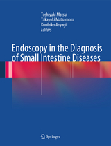
Endoscopy in the Diagnosis of Small Intestine Diseases
Springer Verlag, Japan
978-4-431-54351-0 (ISBN)
From a practical point of view, it is important to observe endoscopic pictures first, then to compare the images of other modalities, and finally to compare macroscopic pictures of resected specimens. For that reason, a large number of well-depicted examples of small intestinal lesions were assembled to clarify differences among small intestinal lesions that appear to exhibit similar findings and morphologies.
Comparisons with radiographic findings comprise another important element in diagnosis. There are limitations in endoscopic observations of gross lesions of the small intestine, with its many convolutions. In Japan, many institutions still practice double-contrast imaging, which provides beautiful results. Because a single disorder may exhibit variations, this volume includes multiple depictions of the same disorders. Also included are lesions in active and inactive phases, as both appearances are highly likely to be encountered simultaneously in clinical practice. The number of illustrated findings therefore has been limited to strictly selected cases.
1, Toshiyuki Matsui, MD, Ph D, Professor, Department of Gastroenterology, Fukuoka University Chikushi Hospital, 2, Takayuki Matsumoto, MD, PhD, Lecturer, Department of Internal Medicine, Kyushu University 3, Kunihiko Aoyagi, MD, PhD, Clinical Professor, Department of Gastroenterology, Fukuoka University
Part 1. General Considerations.- Chapter 1. Diagnostic Process for Small Intestinal Disease.- Chapter 2. Small Intestinal Radiography.- Chapter 3. Capsule Endoscopy.- Chapter 4. Double-Balloon Endoscopy.- Part 2. Specific Findings of Small Intestinal Lesions.- Chapter 5. Protruded Lesions.- Chapter 6. Submucosal Elevations.- Chapter 7. Ulcerative Lesions.- Chapter 8. Aphthous Lesions.- Chapter 9. Stenotic Lesions.- Chapter 10. Hemorrhagic Lesions.- Chapter 11. Diffuse Lesions.- Chapter 12. Reddish Lesions.- Chapter 13. Edematous Lesions.- Chapter 14. Case presentations: Flat / Small protrusions.- Chapter 15. Case Presentations: Depressions.- Chapter 16. Case Presentations: Protrusions of Submucosal Elevations.- Chapter 17. Case presentations: Protrusion with Ulcer.- Chapter 18. Case Presentations: Multiple Protrusions.- Chapter 19. Case Presentations: Ulcers.- Chapter 20. Case Presentations: Stenosis.- Chapter 21. Hemorrhagic Lesions.- Chapter 22. Reddened Lesions.- Chapter 23. Edematous Lesions.- Chapter 24. Erosive Lesions.- Chapter 25. Diffuse Granular or Diffuse Coarse Mucosal Lesions.- Chapter 26. Whitish Multi-Nodular Lesions.- Chapter 27. Intraluminal Growth.- Part 3. Basic Knowledge and Classification.- Chapter 28. On Tumors.- Chapter 29. On Inflammation
| Zusatzinfo | 245 Illustrations, color; 94 Illustrations, black and white; XV, 283 p. 339 illus., 245 illus. in color. |
|---|---|
| Verlagsort | Tokyo |
| Sprache | englisch |
| Maße | 210 x 279 mm |
| Themenwelt | Medizinische Fachgebiete ► Chirurgie ► Viszeralchirurgie |
| Medizinische Fachgebiete ► Innere Medizin ► Gastroenterologie | |
| Medizin / Pharmazie ► Medizinische Fachgebiete ► Onkologie | |
| Schlagworte | Capsule endoscopy • Double-balloon endoscopy • Small Intestine |
| ISBN-10 | 4-431-54351-1 / 4431543511 |
| ISBN-13 | 978-4-431-54351-0 / 9784431543510 |
| Zustand | Neuware |
| Haben Sie eine Frage zum Produkt? |
aus dem Bereich


