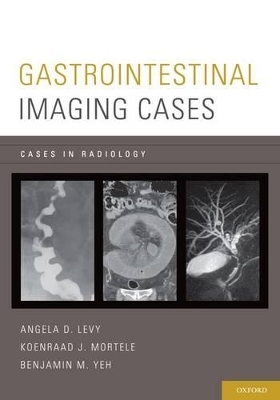
Gastrointestinal Imaging Cases
Seiten
2013
Oxford University Press Inc (Verlag)
978-0-19-975943-9 (ISBN)
Oxford University Press Inc (Verlag)
978-0-19-975943-9 (ISBN)
Gastrointestinal Imaging Cases features 171 unique cases that examine the spectrum of clinical gastrointestinal issues, including both benign and malignant diseases.
Offering 171 unique cases, Gastrointestinal Imaging Cases features over 700 high-quality images. The clinically relevant cases cover both benign and malignant conditions and are grouped into 12 sections organized by the parts of the Gastrointestinal System including: the Pharynx and Esophagus, Stomach, Duodenum, Small Intestine, Appendix, Colon, Rectum, and Anus, Liver, Gallbladder, Bile Ducts, Pancreas, Spleen, and Mesentery and Peritoneum. Within each section, the cases appear in a random order to facilitate the self-assessment experience of reading cases as unknowns. Each case is complete with pertinent findings, differential diagnoses, management recommendations, teaching points, and suggested further reading. This comprehensive, yet easy-to-follow, casebook is an ideal tool for the resident preparing for the boards, the fellow for recertification exams, or the radiologist in need of a quick review.
Offering 171 unique cases, Gastrointestinal Imaging Cases features over 700 high-quality images. The clinically relevant cases cover both benign and malignant conditions and are grouped into 12 sections organized by the parts of the Gastrointestinal System including: the Pharynx and Esophagus, Stomach, Duodenum, Small Intestine, Appendix, Colon, Rectum, and Anus, Liver, Gallbladder, Bile Ducts, Pancreas, Spleen, and Mesentery and Peritoneum. Within each section, the cases appear in a random order to facilitate the self-assessment experience of reading cases as unknowns. Each case is complete with pertinent findings, differential diagnoses, management recommendations, teaching points, and suggested further reading. This comprehensive, yet easy-to-follow, casebook is an ideal tool for the resident preparing for the boards, the fellow for recertification exams, or the radiologist in need of a quick review.
ADL: Professor of Radiology, Georgetown University Medical Center, Washington, DC. KJM: Associate Professor of Radiology, Harvard Medical School, Boston, Massachusetts. BMY: Professor of Radiology, University of California, San Francisco, California.
Contents ; Contributors ; Section 1. Pharynx and Esophagus ; Section 2. Stomach ; Section 3. Duodenum ; Section 4. Small Intestine ; Section 5. Appendix ; Section 6. Colon, Rectum, and Anus ; Section 7. Liver ; Section 8. Gallbladder ; Section 9. Bile Ducts ; Section 10. Pancreas ; Section 11. Spleen ; Section 12. Mesentery and Peritoneum ; Index of Cases ; Index
| Erscheint lt. Verlag | 19.9.2013 |
|---|---|
| Reihe/Serie | Cases In Radiology |
| Zusatzinfo | 728 illustrations |
| Verlagsort | New York |
| Sprache | englisch |
| Maße | 254 x 178 mm |
| Gewicht | 1100 g |
| Themenwelt | Medizinische Fachgebiete ► Innere Medizin ► Gastroenterologie |
| Medizinische Fachgebiete ► Radiologie / Bildgebende Verfahren ► Radiologie | |
| ISBN-10 | 0-19-975943-X / 019975943X |
| ISBN-13 | 978-0-19-975943-9 / 9780199759439 |
| Zustand | Neuware |
| Haben Sie eine Frage zum Produkt? |
Mehr entdecken
aus dem Bereich
aus dem Bereich
Buch | Softcover (2024)
Urban & Fischer in Elsevier (Verlag)
59,00 €


