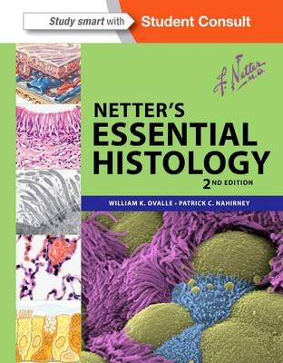
Netter's Essential Histology
with Student Consult Access
Seiten
2013
|
2nd Revised edition
Saunders (Verlag)
978-1-4557-0631-0 (ISBN)
Saunders (Verlag)
978-1-4557-0631-0 (ISBN)
- Titel erscheint in neuer Auflage
- Artikel merken
Zu diesem Artikel existiert eine Nachauflage
Integrates gross anatomy and embryology with classic histology slides and scanning electron microscopy to give you a visual understanding of essential histology. This book utilizes a variety of visual elements - including Netter illustrations and light and electron micrographs - to teach you the histologic concepts and their clinical relevance.
Netter's Essential Histology integrates gross anatomy and embryology with classic histology slides and cutting-edge scanning electron microscopy to give you a rich visual understanding of this complex subject. This histology textbook-atlas has a strong anatomy foundation and utilizes a variety of visual elements - including Netter illustrations and light and electron micrographs - to teach you the most indispensable histologic concepts and their clinical relevance. Excellent as both a reference and a review, Netter's Essential Histology will serve you well at any stage of your healthcare career.
Gain a rich understanding of this vital subject through the succinct explanatory histology text.
Learn to recognize both normal and diseased structures at the microscopic level with the aid of succinct explanatory text as well as numerous clinical boxes.
Access the entire contents and ancillary components online at Student Consult, view images and histology slides at different magnifications, and watch new narrated video overviews of each chapter.
Take your learning one step further with the purchase of Netter's Histology Flash Cards (sold separately), designed to reinforce your understanding of how the human body works in health as well as illness and injury.
Thoroughly comprehend how function is linked to structure through brand-new electron micrographs, many of which have been enhanced and colorized to show ultra-structures in 3D.
Section 1: CELL AND TISSUES
1.The Cell
2.Epithelium and Exocrine Glands
3.Connective Tissue
4.Muscle Tissue
5.Nervous Tissue
6.Cartilage and Bone
7.Blood and Bone Marrow
Section 2: SYSTEMS
8.Cardiovascular System
9.Lymphoid System
10.Endocrine System
11.Integumentary System
12.Upper Digestive System
13.Lower Digestive System
14.Liver, Gallbladder, and Exocrine Pancreas
15.Respiratory System
16.Urinary System
17.Male Reproductive System
18.Female Reproductive System
19.Eye and Adnexa
20.Special Senses
| Reihe/Serie | Netter Basic Science |
|---|---|
| Zusatzinfo | Approx. 500 illustrations (500 in full color) |
| Verlagsort | Philadelphia |
| Sprache | englisch |
| Maße | 216 x 276 mm |
| Themenwelt | Studium ► 1. Studienabschnitt (Vorklinik) ► Histologie / Embryologie |
| ISBN-10 | 1-4557-0631-0 / 1455706310 |
| ISBN-13 | 978-1-4557-0631-0 / 9781455706310 |
| Zustand | Neuware |
| Haben Sie eine Frage zum Produkt? |
Mehr entdecken
aus dem Bereich
aus dem Bereich
Zytologie, Histologie und mikroskopische Anatomie
Buch | Hardcover (2022)
Urban & Fischer in Elsevier (Verlag)
54,00 €


