Learning Chest Imaging
Springer Berlin (Verlag)
978-3-642-34146-5 (ISBN)
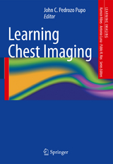
Radiology of the thorax forms an indispensable element of the basic diagnostic process for many conditions and is of key importance in a variety of medical disciplines. This user-friendly book provides an overview of the imaging techniques used in chest radiology and presents numerous instructive case-based images with accompanying explanatory text. A wide range of clinical conditions and circumstances are covered with the aim of enabling the reader to confidently interpret chest images by correctly identifying structures of interest and the causes of abnormalities. This book, which will be an invaluable learning tool, forms part of the Learning Imaging series for medical students, residents, less experienced radiologists, and other medical staff.
Pleural and Pleural Space.- Diaphragm.- Mediastinum and Pulmonary Circulation.- Air Space, and Bronchi I.- Air Space, and Bronchi II.- Trachea and Airway.- Heart and Great Vessels.- Chest Wall and Soft Tissues.- Pulmonary Interstitium.- Medical Device, and Monitoring of the Chest.
From the book reviews:
"This text is part of the 'Learning Imaging' series aimed at medical students, residents, less-experienced radiologists, and other clinicians. ... this book is a good overview of chest imaging. ... For the price and the ease of reading, with good quality images and a comprehensive further reading list, this is a good textbook for the intended audience." (Catherine Jones, Radiology, Vol. 271 (2), May, 2014)
"This book provides an easy to read overview of chest radiography and CT. ... Each of the ten chapters covers a specific anatomical region of the chest and contains a number of case studies illustrating a wide variety of pathologies. ... this book is aimed at medical students, junior radiologists and non-specialist medical staff, and as such is a reasonable introductory text." (Gary Culpan, RAD Magazine, April, 2014)
| Erscheint lt. Verlag | 19.3.2013 |
|---|---|
| Reihe/Serie | Learning Imaging |
| Zusatzinfo | XIV, 242 p. 411 illus., 51 illus. in color. |
| Verlagsort | Berlin |
| Sprache | englisch |
| Maße | 178 x 254 mm |
| Gewicht | 570 g |
| Themenwelt | Medizinische Fachgebiete ► Innere Medizin ► Pneumologie |
| Medizinische Fachgebiete ► Radiologie / Bildgebende Verfahren ► Radiologie | |
| Schlagworte | Bildgebende Verfahren (Medizin) • Brust • Chest Imaging • Chest Radiograph • pneumology • Radiology • Respiratory Diseases |
| ISBN-10 | 3-642-34146-2 / 3642341462 |
| ISBN-13 | 978-3-642-34146-5 / 9783642341465 |
| Zustand | Neuware |
| Informationen gemäß Produktsicherheitsverordnung (GPSR) | |
| Haben Sie eine Frage zum Produkt? |
aus dem Bereich