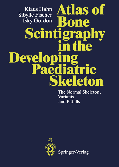
Atlas of Bone Scintigraphy in the Developing Paediatric Skeleton
Springer Berlin (Verlag)
978-3-642-84947-3 (ISBN)
Radioisotope bone scans of the paediatric skeleton have been undertaken in the Departments of Radiology and Nuclear Medicine on a daily basis. Indications for bone scintigraphy include infection, trauma, primary benign bone tumours, as well as malignancy. Other conditions such as avascular necrosis and certain dysplasias also warrant a bone scan. When faced with a child who is symptomatic, but in whom the diagnosis is uncertain, eg. the child with a limp of backache, will require a bone scan to exclude the skeleton as the source of the symptoms. For Departments where paediatric bone scans are carried out infrequently this Atlas will provide a crucial reference for the Radiologist, Nuclear Medicine Physician and Orthopaedic Surgeon, to be able to compare any particular paediatric bone scan with the variations of normality as displayed in the Atlas. Important advice is given to ensure high quality bone scan images which will allow better differentiation between normality and abnormality. This Atlas should be on the shelf of any department which undertakes bone scintigraphy in children, expecially if this is done on an irregular basis.
Pastor Klaus Hahn ist Pädagoge und langjähriger Redaktionsleiter und Herausgeber der Schriftenreihe KU Praxis. Er ist Dozent am Predigerseminar in Bad Kreuznach/Wuppertal.
Sibylle Fischer, BA Pädagogik der Frühen Kindheit, Fachwirtin für Kitas, gibt Fortbildungen für Erzieherinnen und arbeitet als wissenschaftliche Mitarbeiterin an der Evangelischen Hochschule Freiburg.
1 Age 0- 6 Months.- 2 Age 6-12 Months.- 3 Age 1- 2 Years.- 4 Age 2- 3 Years.- 5 Age 3- 4 Years.- 6 Age 4- 5 Years.- 7 Age 5- 6 Years.- 8 Age 6- 7 Years.- 9 Age 7- 8 Years.- 10 Age 8- 9 Years.- 11 Age 9-10 Years.- 12 Age 10-11 Years.- 13 Age 11-12 Years.- 14 Age 12-13 Years.- 15 Age 13-14 Years.- 16 Age 14-15 Years.- 17 Age 15-17 Years.- 18 Age 17-22 Years.- 19 Knees.- 20 Hips.
"This is an excellent reference and a good addition to the imaging libraries of nuclear medicine departments and practices that occasionally image growing children." (Journal of Nuclear Medicine)
| Erscheint lt. Verlag | 15.12.2011 |
|---|---|
| Mitarbeit |
Assistent: J. Guillet, A. Piepsz, I. Roca, M. Wioland |
| Vorwort | D.L. Gilday |
| Zusatzinfo | VIII, 316 p. 283 illus. With 1 Falttafel. |
| Verlagsort | Berlin |
| Sprache | englisch |
| Maße | 193 x 270 mm |
| Gewicht | 712 g |
| Themenwelt | Medizinische Fachgebiete ► Chirurgie ► Unfallchirurgie / Orthopädie |
| Medizin / Pharmazie ► Medizinische Fachgebiete ► Pädiatrie | |
| Medizinische Fachgebiete ► Radiologie / Bildgebende Verfahren ► Nuklearmedizin | |
| Schlagworte | Bone • Diagnosis • Infection • Knochenszintigraphie • Nuclear Medicine • Pädiatrie • Paediatrics • Pediatrics • Radio-isotope Bones • Radiology • Scintigraphy • Trauma • Tumor |
| ISBN-10 | 3-642-84947-4 / 3642849474 |
| ISBN-13 | 978-3-642-84947-3 / 9783642849473 |
| Zustand | Neuware |
| Haben Sie eine Frage zum Produkt? |
aus dem Bereich


