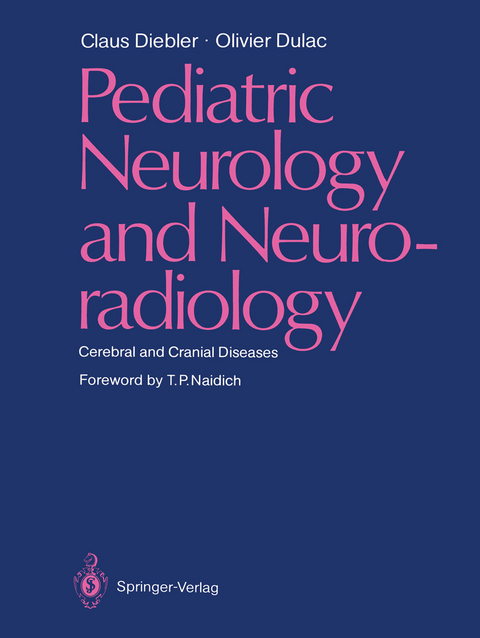
Pediatric Neurology and Neuroradiology
Springer Berlin (Verlag)
978-3-642-70380-5 (ISBN)
1 Cerebral and Cranial Malformations.- 1.1 Malformations of the Corpus Callosum.- 1.2 Prosencephaly: Arhinencephaly.- 1.3 Absence of the Septum Pellucidum.- 1.4 Malformations of the Cerebral Cortex.- 1.5 Congenital Obstruction of the Aqueduct of Sylvius.- 1.6 Cleland-Chiari Malformation.- 1.7 Dandy-Walker Syndrome.- 1.8 Cerebellar Hypoplasia.- 1.9 Cephaloceles.- 1.10 Malformative Intracranial Cysts.- 1.11 Hamartoma of the Tuber Cinereum.- 1.12 Benign External Hydrocephalus.- 1.13 Primary Megalencephaly.- 2 Neurocutaneous Syndromes.- 2.1 Neurofibromatosis.- 2.2 Tuberous Sclerosis.- 2.3 Sturge-Weber Syndrome.- 2.4 Incontinentia Pigmenti.- 2.5 Nevus Linearis Sebaceus Syndrome.- 2.6 Encephalocraniocutaneous Lipomatosis.- 2.7 Hypomelanosis of Itô (Incontinentia Pigmenti Achromians).- 2.8 Neurocutaneous Melanosis.- 2.9 Nevoid Basal Cell Carcinoma Syndrome.- 2.10 Facial Nevi Associated with Anomalous Venous Return and Hydrocephalus.- 3 Inherited Metabolic Diseases.- 3.1 Diseases with Basal Ganglia Lesions.- 3.2 Poliodystrophies.- 3.3 Infantile Neuroaxonal Dystrophy.- 3.4 Lafora's Disease.- 3.5 Leukodystrophies.- 3.6 Kearns-Sayre Syndrome.- 3.7 Mucopolysaccharidosis.- 3.8 Ataxia-Telangiectasia.- 4 Infectious Diseases of the Central Nervous System.- Bacterial Infections.- 4.1 Neonatal Leptomeningitis.- 4.2 Bacterial Leptomeningitis in Infancy and Childhood.- 4.3 Intracranial Tuberculosis.- 4.4 Intracranial Suppuration.- Viral Infections and Diseases of Presumed Viral Origin.- 4.5 Viral Encephalitis.- Parasitic Diseases.- 4.6 Toxoplasmosis.- 4.7 Cerebral Hydatid Cyst.- 4.8 Cysticercosis.- 5 Vascular Disorders.- 5.1 Prenatal and Perinatal Cerebral Lesions of Circulatory Origin.- 5.2 Postnatal Vascular Obstruction.- 5.3 Cranial and Cerebral Vascular Malformations.- 6Intracranial Tumors.- 6.1 Posterior Fossa Tumors.- 6.2 Tumors of the Region of the Third Ventricle and the Region of the Sella Turcica.- 6.3 Tumors of the Cerebral Hemispheres.- 6.4 Orbital Tumors.- 7 Cranial Trauma.- 7.1 Physiopathology of the Cerebral Lesions.- 7.2 Clinical and Radiologic Aspects of Head Trauma in Infancy and Childhood.- 8 Miscellaneous.- 8.1 Osseous Dysplasias.- 8.2 Histiocytosis X.- 8.3 Iatrogenic Diseases.- 8.4 Radionecrosis.- 8.5 Review of Various Symptoms and Syndromes in Infancy and Childhood.
| Erscheint lt. Verlag | 17.11.2011 |
|---|---|
| Vorwort | T.P. Naidich |
| Zusatzinfo | XV, 408 p. |
| Verlagsort | Berlin |
| Sprache | englisch |
| Maße | 210 x 280 mm |
| Gewicht | 1039 g |
| Themenwelt | Medizin / Pharmazie ► Medizinische Fachgebiete ► Neurologie |
| Medizin / Pharmazie ► Medizinische Fachgebiete ► Pädiatrie | |
| Medizinische Fachgebiete ► Radiologie / Bildgebende Verfahren ► Radiologie | |
| Schlagworte | Computed tomography (CT) • Diagnosis • Kinderheilkunde • Neurology • Neuroradiologie • Radiology |
| ISBN-10 | 3-642-70380-1 / 3642703801 |
| ISBN-13 | 978-3-642-70380-5 / 9783642703805 |
| Zustand | Neuware |
| Informationen gemäß Produktsicherheitsverordnung (GPSR) | |
| Haben Sie eine Frage zum Produkt? |
aus dem Bereich


