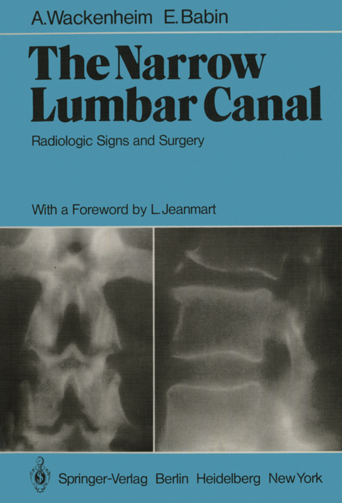
The Narrow Lumbar Canal
Springer Berlin (Verlag)
978-3-642-67349-8 (ISBN)
1. Radiology of the Narrow Lumbar Canal.- 1.1 History.- 1.2 Terminology.- 1.3 Anatomy.- 1.4 Clinical Data.- 1.5 Radiologic Techniques and Their Indications.- 1.6 Radiologic Signs of Lumbar Canal Narrowness.- 1.7 Nosology.- Figures 1-7.- 2. Plain X-Ray Diagnosis of Developmental Narrow Lumbar Canal.- 2.1 Technique.- 2.2 Findings: To Measure or Not to Measure? That is Not the Question.- 2.3 Requirements for Reliable Measurements and Pitfalls.- 2.4 Radiologic Features of the Narrow Lumbar Canal Without Contrast Medium.- 2.5 Findings on Lateral Projection.- 2.6 Various Types of Developmental Stenosis.- 2.7 Correlation Between Surgical and Radiologic Reports.- 2.8 Narrow Lumbar Canal and Associated Diseases.- Figures 8-14.- 3. Interapophysolaminar Spaces (IALS) of the Lumbar Spine and Their Utility in the Diagnosis of Narrow Lumbar Canal.- 3.1 Introduction.- 3.2 Material and Methods.- 3.3 Results.- 3.4 Conclusion.- Figures 15-18.- 4. Myelographic Signs of Narrow Lumbar Canal.- 4.1 Technical Particularities.- 4.2 Limits of the LM as an Investigation of the Narrow Lumbar Canals.- 4.3 LM Anomalies.- Figures 19-28.- 5. Gas Myelography in Verbiest's Developmental Spinal Canal Stenosis.- 5.1 Symptomatology.- 5.2 Radiologic Examination.- 5.3 Clinical Forms of Idiopathic Developmental Spinal Stenosis.- Figures 29-42.- 6. Phlebographic Signs of the Narrow Lumbar CanaL.- 6.1 Physiopathology of the Venous Compression.- 6.2 Phlebographic Signs of Narrow Lumbar Canal.- Figures 43-47.- 7. Narrow Lumbar Canal by Postoperative Epidural Lesions.- 7.1 Radiculosaccographic Semeiology of Epidural Scarring.- 7.2 Phlebographic Semeiology of Epidural Scarring.- 7.3 Surgical Findings.- 7.4 Clinical Aspects.- 7.5 Physiopathology.- 7.6 Conclusion.- Figures 48-60.- 8. SpinalPhlebography in the Stenosis of the Lumbar Canal.- Figures 61-72.- 9. Computerized Tomography in Lumbar Spinal Stenosis.- 9.1 Material and Methods.- 9.2 Results.- 9.3 Conclusion.- Figures 73-84.- 10. Lumbar Spinal Stenosis.- 10.1 Etiology.- 10.2 Symptomatology.- 10.3 Treatment.- Figures 85-98.- 11. Narrow Radicular Canal.- 11.1 Nosologic Importance of the Narrow Radicular Canal with Regard to the Narrow Lumbar Canal.- 11.2 Anatomy of the Radicular Canal.- 11.3 Etiologies.- 11.4 Symptomatology of the Narrow Radicular Canal.- 11.5 Radiologic Findings.- 11.6 Surgical Procedures.- 11.7 Conclusion.- Figures 99-108.- 12. Stenosis of the Bony Lumbar Vertebral Canal.- 12.1 Introduction.- 12.2 Historical Review: Evolution of the Idea.- 12.3 Nomenclature.- 12.4 Classification of the Types of Stenoses of the Lumbar Vertebral Canal.- 12.5 Semiological Aspects.- Figures 125-127.- 12.6 Surgical Treatment and Results.- 13. Cheirolumbar Dysostosis: Developmental Brachycheiry and Narrowness of the Lumbar Canal.- Figures 128-139.- References.- Author Index.
| Erscheint lt. Verlag | 9.12.2011 |
|---|---|
| Einführung | L. Jeanmart |
| Zusatzinfo | XIV, 172 p. |
| Verlagsort | Berlin |
| Sprache | englisch |
| Maße | 210 x 280 mm |
| Gewicht | 483 g |
| Themenwelt | Medizinische Fachgebiete ► Chirurgie ► Neurochirurgie |
| Medizinische Fachgebiete ► Radiologie / Bildgebende Verfahren ► Radiologie | |
| Schlagworte | Lendenwirbelsäulenerkrankung • Medizinische Radiologie • Rückenmarkschirurgie • spinal cord • spine • Surgery • Vertebral column • Wirbelsäulenerkrankung |
| ISBN-10 | 3-642-67349-X / 364267349X |
| ISBN-13 | 978-3-642-67349-8 / 9783642673498 |
| Zustand | Neuware |
| Haben Sie eine Frage zum Produkt? |
aus dem Bereich


