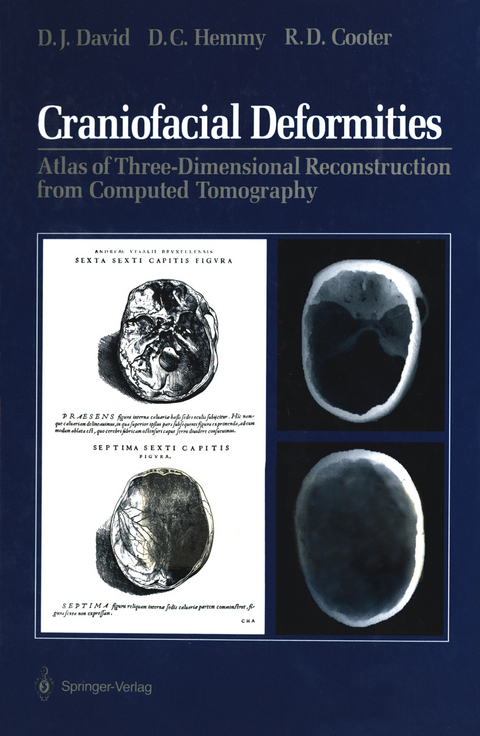
Craniofacial Deformities
Springer-Verlag New York Inc.
978-1-4612-7926-6 (ISBN)
This book has been assembled from the radiographic and photo graphic records of patients presenting to craniofacial units on four continents over 7 years. It is our purpose to illustrate a wide range of craniofacial deformities with the technique of three-dimensional com puted tomography. Many topics are briefly addressed with descriptive text intended to amplify the accompanying images but not to exclude the need for more comprehensive references as recommended in the reading list of each chapter. The ability to generate three-dimensional radiographic images rep resents a successful integration of computed tomography with com puter graphics. Although this technique remains an electronic substi tute for the study of dry skull specimens, it offers a permanent pictorial record of anatomical structures with the opportunity for fu ture interactive data manipulation. It is hoped, therefore, that this work will assist others to gain a more complete understanding of disorders of the craniofacial region. We encourage other surgeons and investigators to examine and employ the techniques used to gather these images but also to ensure that standardized scanning regimens are adapted. The importance of data collection within its full anatomical context was borne out with many of our early studies, which were limited owing to computational con straints. Often an image requirement for surgical intervention is much less than an image necessary for strict scientific inquiry.
1 History of Three-Dimensional Imaging of Craniofacial Disorders.- 2 Three-Dimensional Imaging Techniques.- CT Scanning.- Data Selection.- Image Processing.- Work Station.- Work Station Products.- 3 Normal Skull.- Technique.- 4 Craniosynostoses.- Simple Calvarial Deformities.- Scaphocephaly.- Trigonocephaly.- Turricephaly.- Oxycephaly.- Plagiocephaly.- Craniofacial Syndromes.- Crouzon Syndrome.- Apert Syndrome.- Saethre-Chotzen Syndrome.- Cohen Syndrome.- Pfeiffer Syndrome.- 5 Craniofacial Clefts.- “Tessier” Craniofacial Clefts.- Treacher Collins Syndrome.- Craniofacial Microsomia.- 6 Meningoencephaloceles.- Frontoethmoidal Meningoencephalocele.- Nasofrontal Defect.- Nasoethmoidal Defect.- Nasoorbital Defect.- Basal Meningoencephalocele.- 7 Growth Disorders.- Primary Growth Disorders.- Microorbitism.- Binder Syndrome.- Secondary Growth Disorders.- Romberg Syndrome.- Radiation Effects.- 8 Tumors.- Fibrous Dysplasia.- Neurofibromatosis.- Hemangiomas.- 9 Trauma.- Orbitocranial Fractures.- Midfacial Fractures.- Le Fort I (Guerin) Fracture.- Le Fort II (Pyramidal) Fracture.- Le Fort III Fracture (Cranial Disjunction).- Gunshot Wounds.- Old Trauma.
| Zusatzinfo | X, 147 p. |
|---|---|
| Verlagsort | New York, NY |
| Sprache | englisch |
| Maße | 178 x 254 mm |
| Themenwelt | Medizinische Fachgebiete ► Chirurgie ► Ästhetische und Plastische Chirurgie |
| Medizinische Fachgebiete ► Chirurgie ► Neurochirurgie | |
| Medizin / Pharmazie ► Medizinische Fachgebiete ► HNO-Heilkunde | |
| Medizinische Fachgebiete ► Radiologie / Bildgebende Verfahren ► Radiologie | |
| ISBN-10 | 1-4612-7926-7 / 1461279267 |
| ISBN-13 | 978-1-4612-7926-6 / 9781461279266 |
| Zustand | Neuware |
| Haben Sie eine Frage zum Produkt? |
aus dem Bereich


