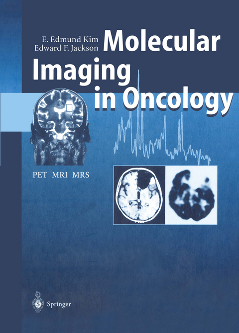
Molecular Imaging in Oncology
Springer Berlin (Verlag)
978-3-642-64163-3 (ISBN)
Principles and Technology.- 1 Principles of Cancer Biology, Biochemistry, Immunology and Pathology.- 2 Imaging Strategies and Perspectives in Oncology.- 3 Magnetic Resonance Imaging: Physical Principles to Advanced Applications.- 4 Magnetic Resonance Spectroscopy: Physical Principles and Applications.- 5 Principles and Instrumentation of Position Emission Tomography.- 6 Radiopharmaceuticals for Tumor Imaging and Magnetic Resonance Imaging Contrast Agents.- 7 Receptor Imaging.- 8 Practical Magnetic Resonance Imaging and Positron Emission Tomography Techniques and Their Artifacts.- Clinical Applications of MRI, MRS and PET.- 9 Lung Cancers.- 10 Breast Cancer.- 11 Gastrointestinal Carcinomas.- 12 Urologic Cancers.- 13 Gynecologic Cancers.- 14 Brain Tumors.- 15 Head and Neck Tumors.- 16 Musculoskeletal Tumors.- 17 Melanoma, Lymphoma and Myeloma.
From the reviews:
"This handbook focuses on the growing impact of molecular imaging in oncology and addresses topics ranging from basic research to clinical applications in the era of evidence-based medicine. ... I highly recommend this book to nuclear physicians, radiologists, oncologists, chemists, physicists, mathematicians, and computer scientists." (E. Edmund Kim, The Journal of Nuclear Medicine, Vol. 54 (6), June, 2013)
| Erscheint lt. Verlag | 30.9.2011 |
|---|---|
| Co-Autor | J. Aoki, H. Baghaei, S. Ilgan, T. Inoue, H. Li, J. Uribe, F.C.L. Wong, W.-H. Wong, D.J. Yang |
| Zusatzinfo | XIII, 290 p. 5 illus. in color. |
| Verlagsort | Berlin |
| Sprache | englisch |
| Maße | 193 x 270 mm |
| Gewicht | 670 g |
| Themenwelt | Medizin / Pharmazie ► Medizinische Fachgebiete ► Onkologie |
| Medizinische Fachgebiete ► Radiologie / Bildgebende Verfahren ► Nuklearmedizin | |
| Medizinische Fachgebiete ► Radiologie / Bildgebende Verfahren ► Radiologie | |
| Schlagworte | brain • Brain Tumors • Breast Cancer • Cancer • Diagnosis • Functional Imaging • Imaging • Lymphoma • Magnetic Resonance Imaging (MRI) • magnetic resonance spectroscopy • melanoma • Metabolic Imaging • Molecular Imaging • MRI • MRS • Pathology • PET • positron emission tomography (PET) • Tomography • Tumor |
| ISBN-10 | 3-642-64163-6 / 3642641636 |
| ISBN-13 | 978-3-642-64163-3 / 9783642641633 |
| Zustand | Neuware |
| Informationen gemäß Produktsicherheitsverordnung (GPSR) | |
| Haben Sie eine Frage zum Produkt? |
aus dem Bereich


