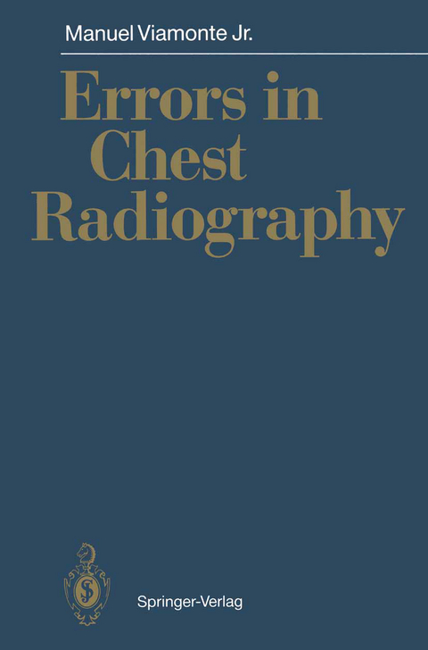
Errors in Chest Radiography
Springer Berlin (Verlag)
978-3-540-52906-4 (ISBN)
Warum entstehen Fehler bei radiologischen Untersuchungen des Brustraums? Der Autor klärt Ursachen und zeigt, daß aus Fehlern gelernt werden kann, um sie in Zukunft zu vermeiden. Jeder Radiologe wird aus dieser Fallsammlung Nutzen ziehen!
Chest radiography is the most commonly perfonned diagnostic radiological exam ination in the United States. More than 80 million chest radiographs are perfonned annually in the United States and this type of radiograph accounts for 30%-50% of the total volume of diagnostic studies. Standard chest radiographic examinations are difficult to optimize, primarily because of the seven-to tenfold greater attenuation of the mediastinum and heart than of the lungs. In order to obtain the best results, we must be able to see with distinct clarity the vascular markings of the lungs, particularly when these are superimposed on the rib cage, the bony structures, and the air-soft tissue interfaces of the mediastinum. The large variation in attenuation caused by the mediastinal structures cannot be recorded routinely on a radiograph with maximum image contrast. The lungs are shaped like a truncated cone. The apices are volumetrically smaller than the bases and are crisscrossed by bony structures (the upper ribs, clavicle, and sometimes the scapula and manubrium of sternum). Often in women, the density of the breasts overlaps the lung bases, and X-rays must therefore traverse more tissue. Despite the cephalocaudal increase in tissue volume, it is possible in most in stances to obtain a balanced density and contrast from the apices to the bases using the high kilovoltage peak (kVp) technique. Optimal image quality demands appro priate resolution and contrast that will pennit the detection of pulmonary opacities and lucencies and of mediastinal and chest wall abnonnalities.
Technical Aspects.- Film-Screen Combination.- Techniques for Scatter Reduction.- Digital Radiography.- Automated Chest Unit.- Summary.- Errors in Chest Radiology.- Errors in Technique.- Errors in Interpretation.- Difficult Areas of Interpretation.- Pitfalls of Improper Technique and of Interpretation.- References.- Atlas.- Air-Soft Tissue Interfaces with the Mediastinum.- Importance of Using the High kVp Technique.- Importance of Proper Centering.- Underexposure.- Overexposure.- Effect of Poor Inspiratory Effort.- Importance of Including a Lateral View.- Importance of Including an Oblique View.- Failure to Interpret Results Correctly.- Difficult Areas to Assess.- Hyperlucency Which is Often Misinterpreted.- Importance of Correct Examination of the Upper Airways.- Importance of Correct Examination of the Bones.- Importance of Correct Examination of the Soft Tissues.- Importance of Correct Examination of the Abdomen.- Importance of Adequate Knowledge of Patient's Clinical History.- Importance of Careful Examination of the Hila.- Learn About Your Patient.- Don't Procrastinate.- Value of the Esophagogram.- Value of Linear Tomography.- Value of Computed Tomography.- Value of Ultrasonography.- Value of Arteriogram.- Value of AMBER Study.
| Erscheint lt. Verlag | 29.1.1991 |
|---|---|
| Zusatzinfo | VI, 135 p. |
| Verlagsort | Berlin |
| Sprache | englisch |
| Maße | 155 x 235 mm |
| Gewicht | 260 g |
| Themenwelt | Medizinische Fachgebiete ► Radiologie / Bildgebende Verfahren ► Radiologie |
| Schlagworte | Bronchial Cancer • Bronchialkrebs • Brust • Chest Radiology • Computed tomography (CT) • Diagnosis in Radiology • Fehldiagnose • Mediastinum • Radiographie • Radiologische • Radiology |
| ISBN-10 | 3-540-52906-3 / 3540529063 |
| ISBN-13 | 978-3-540-52906-4 / 9783540529064 |
| Zustand | Neuware |
| Haben Sie eine Frage zum Produkt? |
aus dem Bereich


