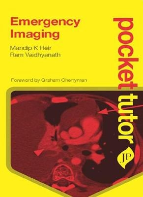
Pocket Tutor Emergency Imaging
JP Medical Ltd (Verlag)
978-1-907816-56-7 (ISBN)
- Titel ist leider vergriffen;
keine Neuauflage - Artikel merken
Titles in the Pocket Tutor series give practical guidance on subjects that medical students and foundation doctors need help with “on the go”, at a highly affordable price that puts them within reach of those rotating through modular courses or working on attachment.
Topics reflect information needs stemming from today’s integrated undergraduate & foundation courses:
Common investigations (ECG, imaging, etc)
Clinical skills (surface anatomy, patient examination, etc.)
Clinical specialties that students perceive as too small to merit a textbook (psychiatry, renal medicine)
Key Points
Highly affordable price and convenient pocket size format – fits in back pocket!
Logical, sequential content: the first principles of emergency imaging, then a guide to understanding a normal image and the building blocks of an abnormal image, before describing specific clinical disorders
Clinical disorders are illustrated by high quality radiographs, ultrasounds, CTs and MRIs, with brief accompanying text that clearly identifies the defining feature of the image
Focuses on the conditions that medical students and foundation doctors are most likely to see and be tested on
Second edition features a new chapter on common emergency cases including chest pain and breathlessness, acute abdominal pain, sudden onset headache, and weight loss and jaundice
Previous edition (9781907816567) published in 2013.
Mandip K Heir MBChB PG Dip MedEd FRCR Consultant Radiologist Ram Vaidhyanath DMRD DNB FRCR EBiHNR Consultant Radiologist, The RCR - Dr PK Ganguli Visiting Professor Both at University Hospitals of Leicester NHS Trust, Leicester, UK
Chapter 1: First principles of emergency imaging
1.1 Imaging modalities
1.2 Use of contrast media
1.3 Investigation requesting and image interpretation
Chapter 2: Understanding normal results
2.1 Plain radiographs
2.2 Ultrasound
2.3 Computed tomography
2.4 Magnetic resonance imaging
Chapter 3: Recognising abnormalities
3.1 Fractures
3.2 Inflammation and abscess
3.3 Effusion
3.4 Haemorrhage
3.5 Thrombosis
3.6 Tumours and mass lesions
3.7 Calcifications
3.8 Foreign bodies
Chapter 4: Gastrointestinal system
4.1 Key radiological anatomy
4.2 Trauma
4.3 Acute inflammation
4.4 Bowel obstruction
4.5 Acute mesenteric ischaemia
4.6 Acute gastrointestinal haemorrhage
Chapter 5: Genitourinary system
5.1 Key radiological anatomy
5.2 Renal trauma
5.3 Bladder trauma
5.4 Urinary tract calculi
5.5 Testicular torsion
5.6 Ovarian torsion
Chapter 6: Chest and vascular disease
6.1 Key radiological anatomy
6.2 Thoracic trauma
6.3 Acute aortic syndrome
6.4 Abdominal aortic aneurysm
6.5 Deep vein thrombosis
6.6 Pulmonary embolism
6.7 Foreign bodies
Chapter 7: Head and neck
7.1 Key radiological anatomy
7.2 Facial trauma
7.3 Orbital trauma
7.4 Orbital infection
7.5 Retropharyngeal abscess
7.6 Foreign bodies
Chapter 8: Neurological imaging
8.1 Key radiological anatomy
8.2 Head injury
8.3 Extradural haemorrhage
8.4 Subdural haemorrhage
8.5 Subarachnoid haemorrhage
8.6 Carotid/vertebral artery dissection
8.7 Stroke
8.8 Cerebral venous thrombosis
8.9 Space-occupying lesions
Chapter 9: Musculoskeletal system
9.1 Key radiological anatomy
9.2 Cervical spine injuries
9.3 Thoracic spine injuries
9.4 Lumbar spine injuries
9.5 Cauda equina compression
9.6 Spondylodiscitis
Chapter 10: Paediatric emergency imaging
10.1 Upper gastrointestinal tract disorders
10.2 Lower gastrointestinal tract disorders
10.3 Musculoskeletal disorders
Chapter 11: Emergency cases
11.1 Chest pain and breathlessness
11.2 Acute abdominal pain
11.3 Low back pain
11.4 Swollen right eye
11.5 Weight loss and jaundice
11.6 Neck pain after a fall
11.7 Collapse
11.8 Sudden onset headache
Index
| Erscheint lt. Verlag | 28.2.2013 |
|---|---|
| Reihe/Serie | Pocket Tutor series |
| Zusatzinfo | 160 Halftones, color |
| Verlagsort | London |
| Sprache | englisch |
| Maße | 113 x 177 mm |
| Themenwelt | Medizin / Pharmazie ► Medizinische Fachgebiete ► Notfallmedizin |
| Medizinische Fachgebiete ► Radiologie / Bildgebende Verfahren ► Radiologie | |
| ISBN-10 | 1-907816-56-9 / 1907816569 |
| ISBN-13 | 978-1-907816-56-7 / 9781907816567 |
| Zustand | Neuware |
| Haben Sie eine Frage zum Produkt? |
aus dem Bereich


