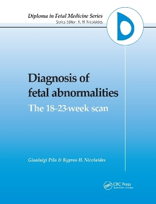
Diagnosis of Fetal Abnormalities
The 18-23-Week Scan
Seiten
1999
Informa Healthcare (Verlag)
978-1-85070-492-8 (ISBN)
Informa Healthcare (Verlag)
978-1-85070-492-8 (ISBN)
A clinician's textbook on using ultrasound as the main diagnostic tool in the prenatal detection of congenital abnormalities. It summarizes the prevalence, aetiology, prenatal sonographic features and prognosis for both common and rare foetal abnormalities.
Ultrasound is the main diagnostic tool in the prenatal detection of congenital abnormalities. The Fetal Medicine Foundation has recognized the importance of this tool by setting up a program of training and certification to help establish high standards of scanning on an international scale. Diagnosis of Fetal Abnormalities: The 18-23-Week Scan provides the basis of learning for the theoretical component of this program. The book is a complete, authoritative clinician's textbook on using ultrasound in the prenatal detection of congenital abnormalities. It summarizes the prevalence, etiology, prenatal sonographic features and prognosis for both common and rare fetal abnormalities.
Ultrasound is the main diagnostic tool in the prenatal detection of congenital abnormalities. The Fetal Medicine Foundation has recognized the importance of this tool by setting up a program of training and certification to help establish high standards of scanning on an international scale. Diagnosis of Fetal Abnormalities: The 18-23-Week Scan provides the basis of learning for the theoretical component of this program. The book is a complete, authoritative clinician's textbook on using ultrasound in the prenatal detection of congenital abnormalities. It summarizes the prevalence, etiology, prenatal sonographic features and prognosis for both common and rare fetal abnormalities.
Gianluigi Pilu, Kypros H. Nicolaides
Introduction. Standard views for examination of the fetus. Central nervous system. Face. Cardiovascular system. Pulmonary abnormalities. Anterior abdominal wall. Gastrointestinal tract. Kidneys and urinary tract. Skeleton. Features of chromosomal defects. Fetal tumors. Hydrops fetalis. Small for gestational age. Abnormalities of the amniotic fluid volume. Appendix I: Risk of major trisomies in relation to maternal age and gestation. Appendix II: Antenatal sonographic findings in skeletal dysplasias. Appendix III: Fetal biometry at 14-40 weeks of gestation. Web sites providing useful information for prenatal diagnosis. Index.
| Erscheint lt. Verlag | 15.6.1999 |
|---|---|
| Zusatzinfo | 50 Line drawings, black and white; 100 Illustrations, black and white |
| Sprache | englisch |
| Maße | 189 x 246 mm |
| Gewicht | 585 g |
| Themenwelt | Medizin / Pharmazie ► Medizinische Fachgebiete ► Gynäkologie / Geburtshilfe |
| Medizinische Fachgebiete ► Radiologie / Bildgebende Verfahren ► Sonographie / Echokardiographie | |
| ISBN-10 | 1-85070-492-9 / 1850704929 |
| ISBN-13 | 978-1-85070-492-8 / 9781850704928 |
| Zustand | Neuware |
| Haben Sie eine Frage zum Produkt? |
Mehr entdecken
aus dem Bereich
aus dem Bereich
Begleitbuch für Sonografiekurse, Klinik und Praxis
Buch | Softcover (2023)
Urban & Fischer in Elsevier (Verlag)
27,00 €
Organbezogene Darstellung von Grund- und Aufbaukurs sowie …
Buch | Hardcover (2020)
Deutscher Ärzteverlag
99,99 €


